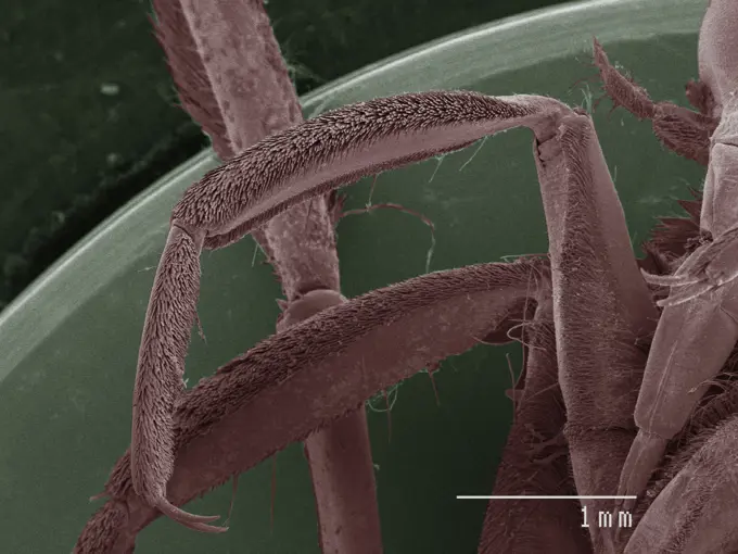
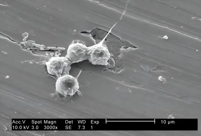
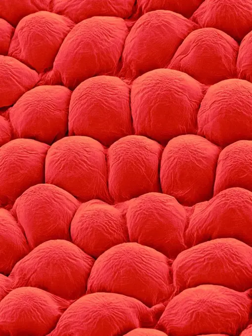
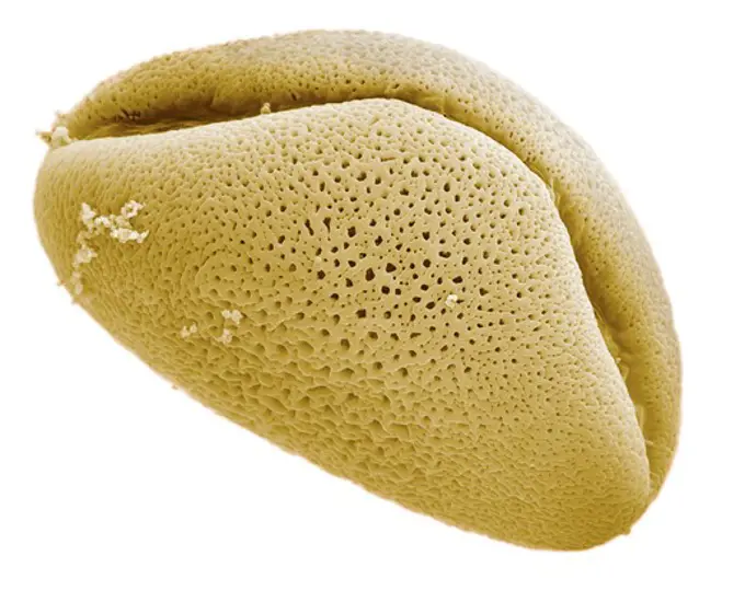
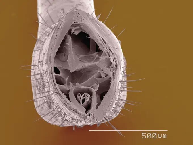
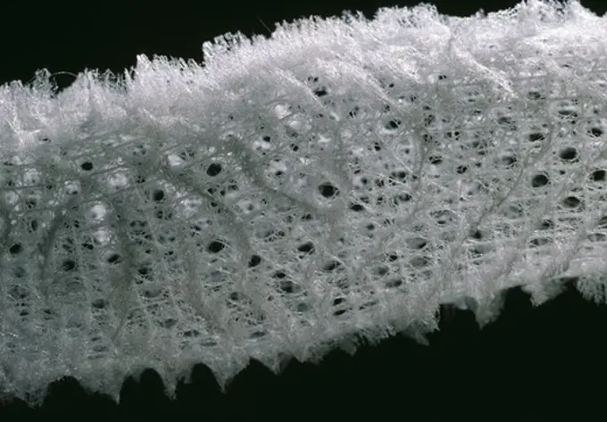
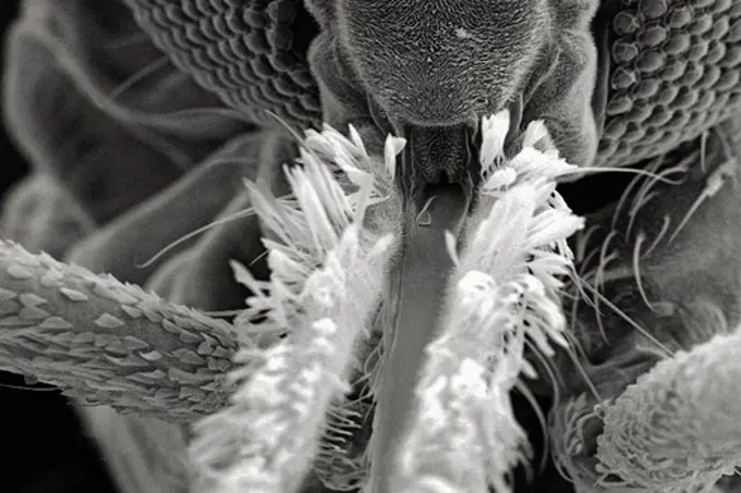
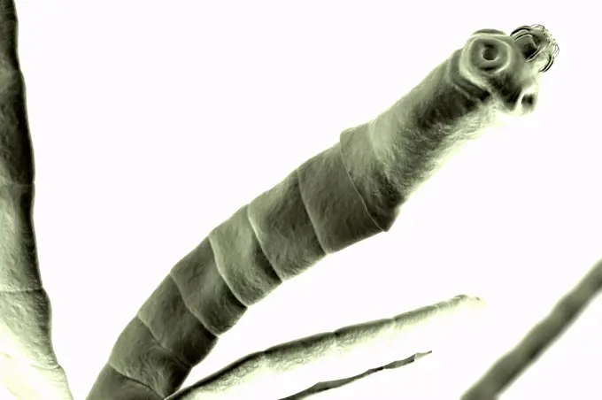
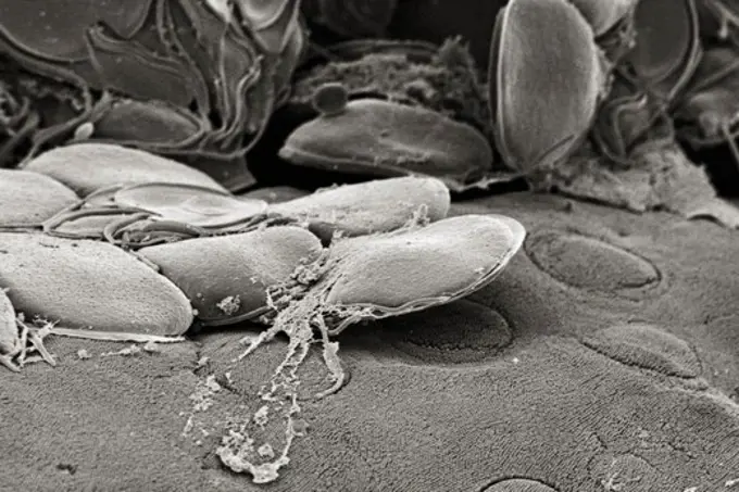
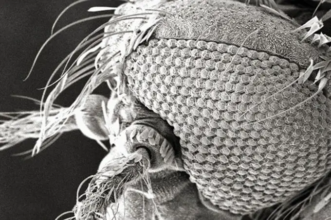
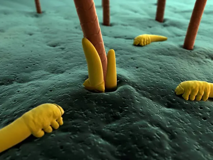
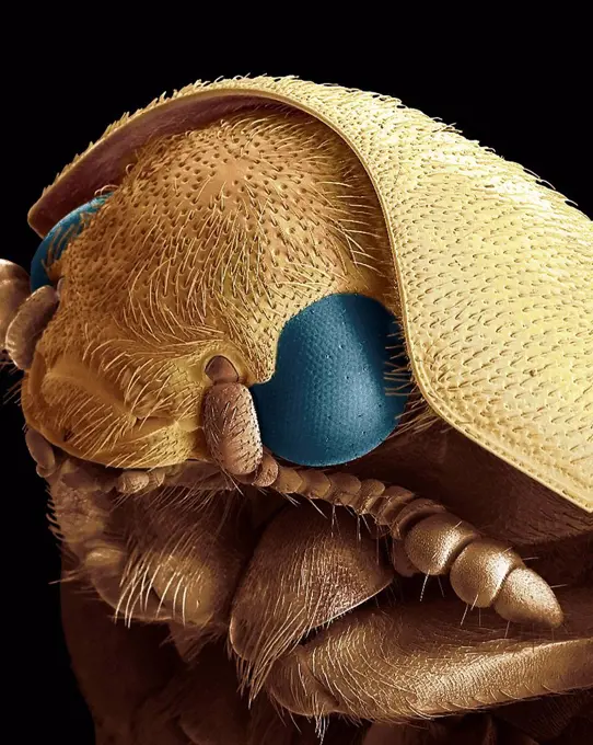
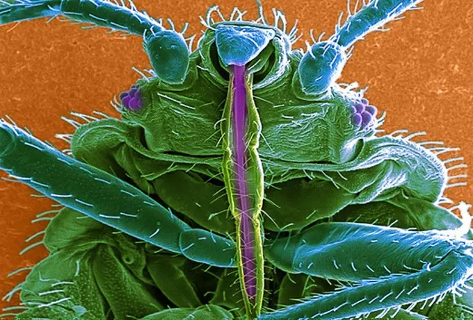
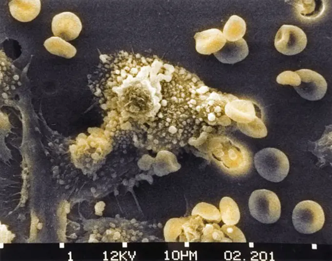
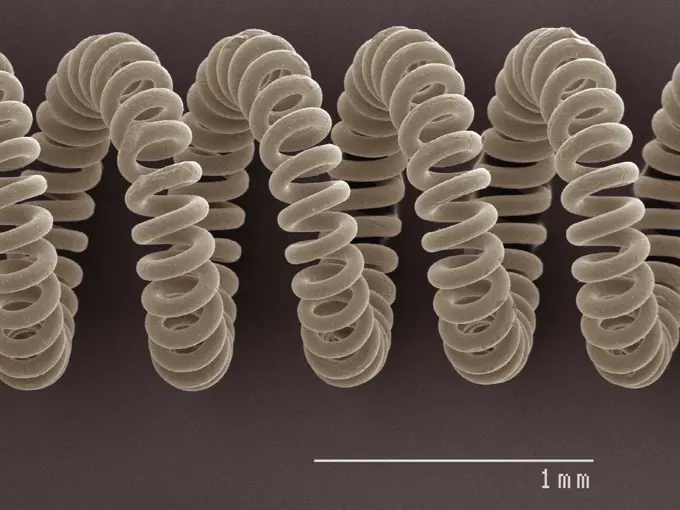
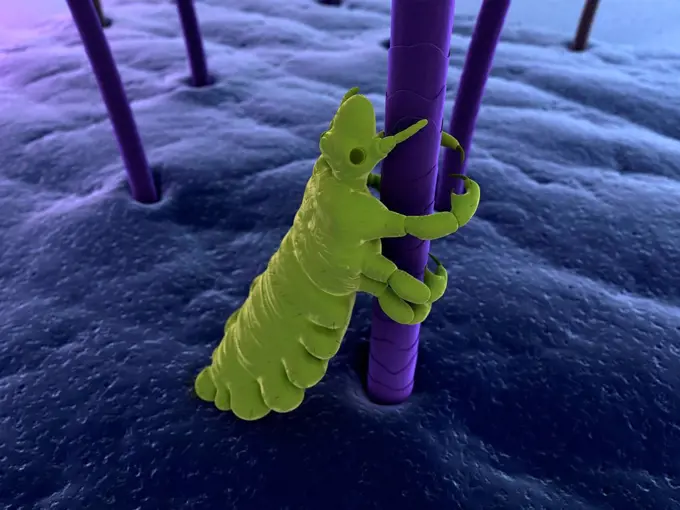
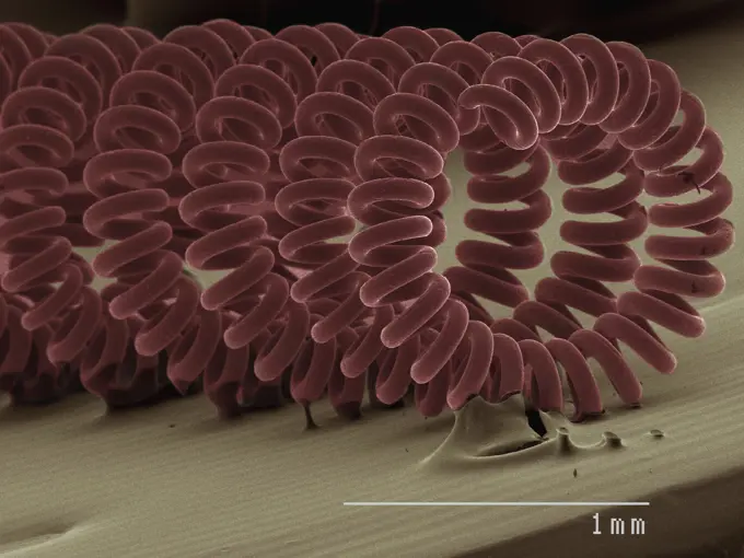
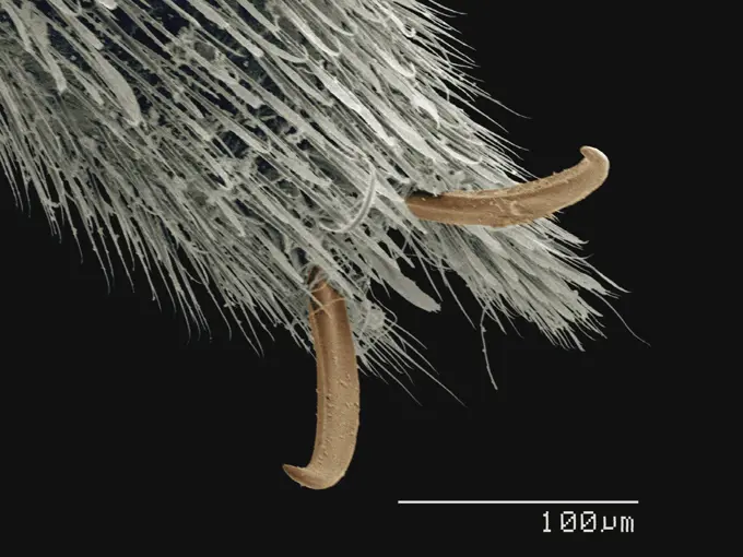
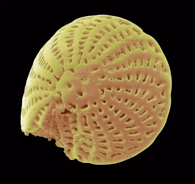
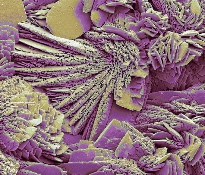
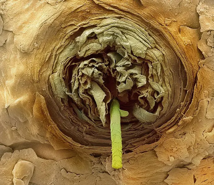
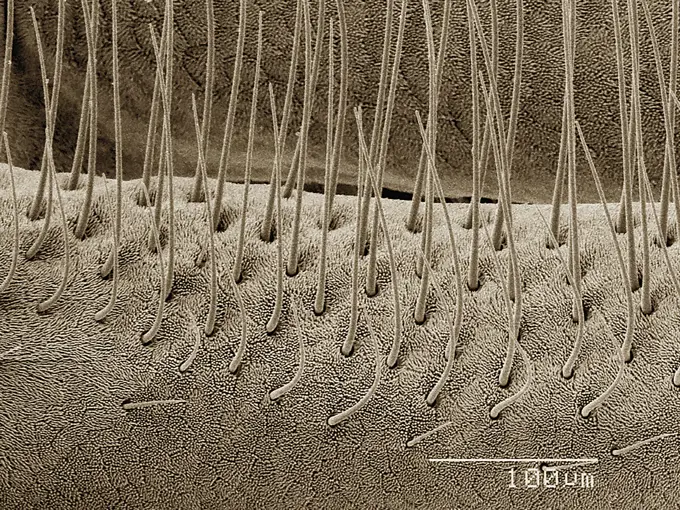
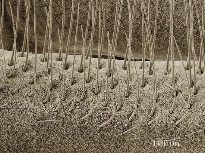
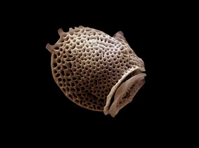
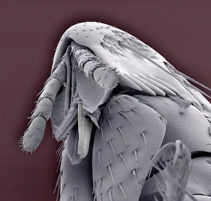
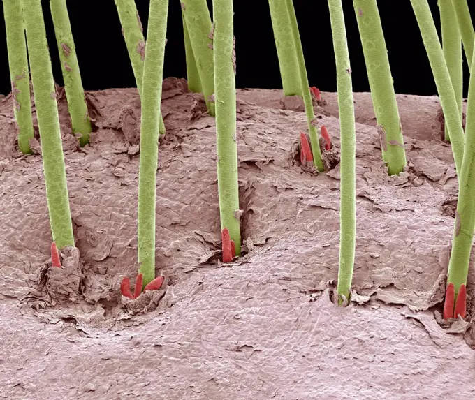
My Boards
View Pictures
  • My Boards
← Science

Microscopic Wonders of Biology

Extreme close-ups of organic structures and insect appendages, showcasing intricate textures and colors using advanced microscopy techniques.

Extreme closeup of insect appendage under the microscope
160 assets in this story
824-57659295
Magnified 3000X, this 2002 scanning electron microscope (SEM) image revealed some of the ultrastructural morphology exhibited by four Hartmannella vermiformis amoeba cysts. The amoeba assumes the cystic stage of its life cycle under harsh conditions such as drought, or when there is an accumulation of toxins in its environment, which can reduce its metabolic needs. Under these circumstances, the protozoan produces a protective coat and goes dormant to wait for better conditions. CDC/Dr Rodney Donlan 2002
4384-221
Scanning Electron Micrograph of Hornet's Leg
4128R-13620157
Papillae on the upper surface of a rose flower petal (Rosa sp), coloured scanning electron micrograph (SEM). Papillae are projections from epidermal c...
1439-57949050
Extreme closeup of insect barbs under an electron microscope
4297-1271
Scanning electron microscopic image of a pollen grain of an Alium
1439-57940187
Closeup view of an intricate microscopic structure
4201-66287
SEM close-up view of a Intertidal Crustacean at 70x magnification, an unidentified species, found on Kihim Beach, Mumbai, India
1439-57943319
Upclose look at an ant head under the microscope
1439-57940183
Closeup of a tiny creature under the microscope
4141-49943
venus's flower-basket sponge, euplectella aspergillum, close detail
4384-411
Microscopic Detail of Mosquito's Head
4378-1200
Microscopic styled visualization of a tapeworm (Cestoda).
1439-57952663
Intricate details of an ant captured in a closeup micrograph
1439-57940162
Closeup view of intricate microstructures
4384-306
Group of Giardia Trophozoites
4384-402
Microscopic Detail of Mosquito's Head
4128R-14506
Eyelash mites Demodex sp., computer artwork.
1439-57952660
Intricate details of a tiny insect magnified under the microscope
4128-111584770
Dermestid beetle. Scanning electron micrograph of a dermestid beetle. Dermestidae are a family of Coleoptera that are commonly referred to as skin bee...
4409-28534314
MINERAL-MALAQUITA PROCEDENTE DEL CONGO.
824-57659290
Magnified 3000X, this 2002 scanning electron microscope (SEM) image revealed part of the ultrastructure exhibited by a number of Hartmannella vermiformis amoeba cysts. The amoeba assumes the cystic stage of its life cycle under harsh conditions such as drought, or when there is an accumulation of toxins in its environment, which can reduce its metabolic needs. Under these circumstances, the protozoan produces a protective coat and goes dormant to wait for better conditions. There is another major life cycle stage that this organism goes through known as the trophozoite stage which is its vegetative phase. At this point, the creature spends time feeding, moving, and reproducing. This free-living protozoan moves in response to chemical cues in its environment by extending pseudopodia or false feet. CDC/Dr Rodney Donlan 2002
4128R-12702
Hoverfly head, coloured scanning electron micrograph SEM. Magnification: x30 when printed at 10 centimetres wide.
4297-1722
Scanning electron micrograph of the ventral surface of a Giardia trophozoite. SEM showing a circular lesion on the intestine (on the left) produced by the ventral adhesive disk of a Giardia protozoan
4128R-11310476
Acrospaera radiolarian. Coloured scanning electron micrograph (SEM) of the shell of an Acrospaera sp. radiolarian. Radiolaria are single-celled protoz...
4128R-9305
Scorpion fly foot, SEM
4297-1136
Scanning electron micrograph revealing the ultrastructural morphology displayed on the ventral surface of a Bedbug (Cimex lectularius)
1899-53513319
Scanning electron micrograph of filamentous Ebola virus particles budding from an infected VERO E6 cell (35,000x magnification). Image captured at the NIAID Integrated Research Facility in Ft. Detrick, Maryland.
4297-1744
SEM image of the Death of a Cancer Cell (step 3 of a 6 step sequence)
1439-57940028
Closeup view of microscopic spiral structures
1439-57940022
Closeup of tiny structures in sepia tones
4128R-15971
Head louse Pediculus humanus capitis, computer artwork.
1439-57940026
Microscopic view of a coiled structure in sepia tones.
1439-57940179
Intricate microstructures resembling an alien landscape
1439-57949043
Closeup of tiny hooks on a surface
4128R-13022178
Foraminiferan. Coloured scanning electron micrograph (SEM) of a foraminiferan microfossil from maldives beach sand. Microfossils' are roughly 0.05 to ...
1439-57940153
Extreme closeup of a chain under a microscope revealing intricate details.
4128-V58557934
4128R-34267
Limescale, coloured scanning electron micrograph (SEM). Limescale, which occurs when hard water is boiled, is composed of calcium or magnesium carbona...
4128R-6044
Eyelash follicle, coloured scanning electron micrograph SEM. Magnification: x400 when printed at 10 centimetres wide.
4297-1691
Scanning electron micrograph depicting a mass of Yersinia pestis bacteria (the cause of bubonic plague) in the foregut of the flea vector
6145-45295498
jsc2021e019394 (5/14/2021) --- Scanning Electron Micrograph images of calcium oxalate microcrystals generated at the University of Washington & Kidney Research Institute. Effects of Microgravity on the Structure and Function of Proximal and Distal Tubule MPS (Kidney Cells-02) uses a 3D kidney cell model or chip to study the effects of microgravity on formation of microcrystals in kidney tubules.Image courtesy of Jacelyn Bain (Kelly Lab) & UW Molecular Analysis Facility.
6145-44607544
Layered Remnant
1773-209589
Ground ivy, x1100 magnification
1916-111284739
A magnified view of its beautiful eye; a special technique was used to remove the ommatidum reflections, leaving an image with SEM like surface detail
1439-57952405
Close-up of cat tongue texture and papillae.
1439-57949086
Closeup view of a gecko's foot under the microscope
6145-46703945
Microscope image of flax. Enlarged 200 times.
4297-1858
Scanning electron microscopic image of Severe Acute Respiratory Syndrome virus particles
4269-27425
Klebsiella pneumoniae. Scanning electron micrograph (SEM) of Klebsiella pneumoniae. K. pneumoniae is a gram-negative, rod-shaped bacterium commonly found in the human gastrointestinal tract. It is responsible for various infections of the urinary tract and lungs (pneumonia), and are often involved in nosocomial infections.
1525-56903810
illustration mushroom, blue, dark blue realistic texture 3D high resolution background fungi
6145-45008545
This scanning electron microscope image shows speroidal features embedded in a layer of iddingsite, a mineral formed by action of water, in a meteorite that came from Mars.
4128R-13928446
Dust mite (Dermatophagoides pteronyssinus), computer illustration. This mite is eight legged. Its rounded, unsegmented body has a few touch-sensitive ...
4128-16171808
Water bear. Coloured scanning electron micrograph (SEM) of a water bear, or tardigrade (phylum Tardigrada). Water bears are small, water-dwelling, seg...
4220-21334522
Scanning Electron Micrograph (SEM): Dinoflagellate, Dinophysis sp;.     Date:
1439-57940018
Closeup view of an object under a scanning electron microscope.
1439-57940133
A closeup view of sepiatoned matches with pink tips arranged in rows.
4384-343
Surface Details of Unidentified Insect's Wing
4128R-10571
Rabbit flea, SEM
4128R-34263
Eyelash mites. Coloured scanning electron micrograph (SEM) of human eyelash mites (Demodex sp.) around hair follicles. Magnification: x35 when printed...
4128R-383
Eyelash follicle, coloured scanning electron micrograph SEM. Magnification: x400 when printed at 10 centimetres wide.
PREVIOUS
of 2
NEXT
2401 S. Ervay, Suite 206
Dallas, Texas 75215
United States
Get Started
Free ResearchMy BoardsMy Cart
For Creators
How To License Your ContentContributor PortalFrame of Mind
Resources
API accessPricing
Contact
+1 866 236 0087help@viewpictures.co.uk Contact form
©2026 View Pictures. All Rights Reserved. -A
This site is protected by reCAPTCHA and Google Privacy Policy and Terms of Service apply.