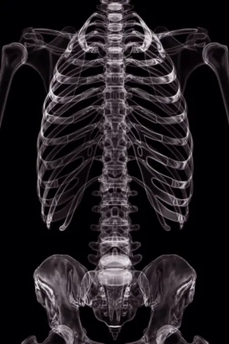
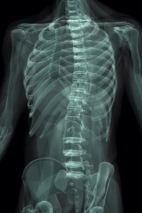
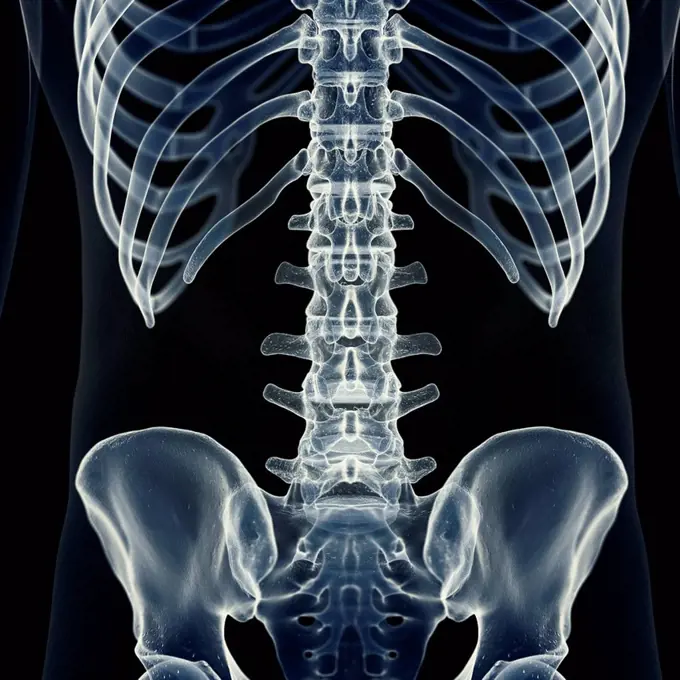
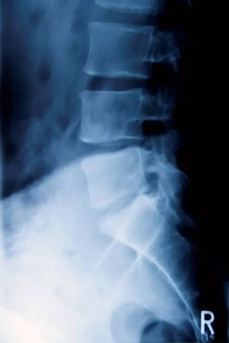
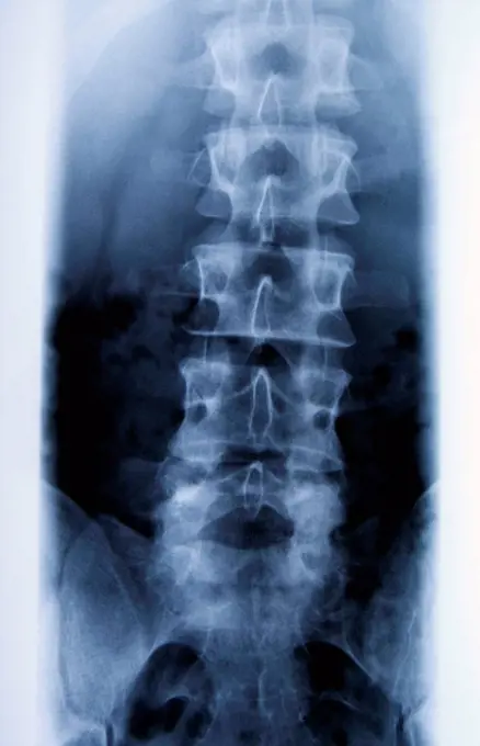
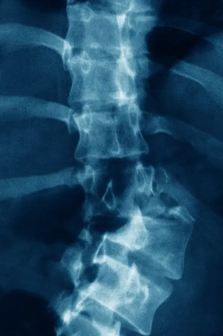
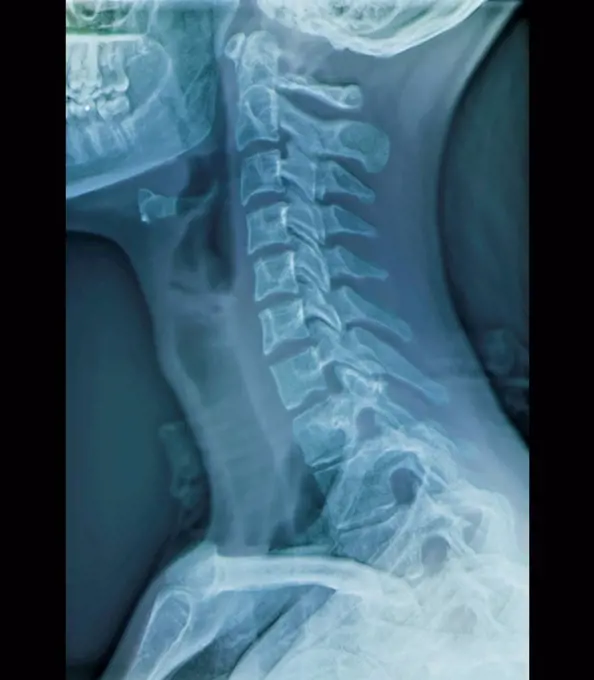
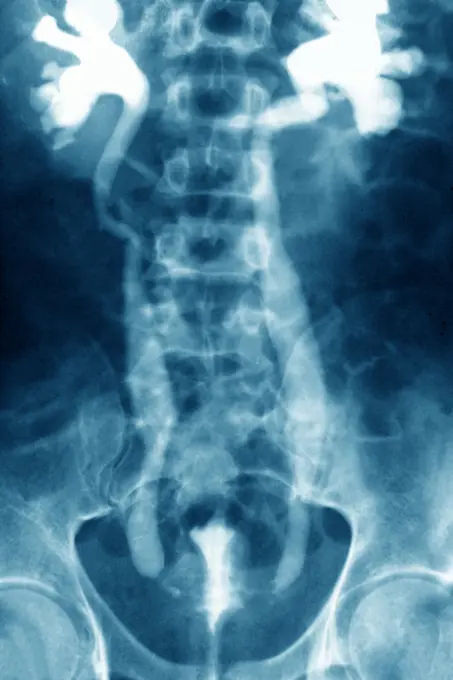
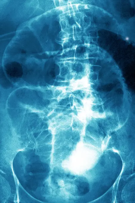
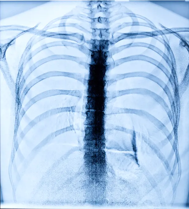
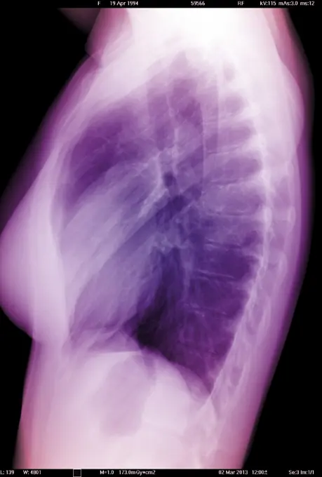
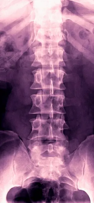
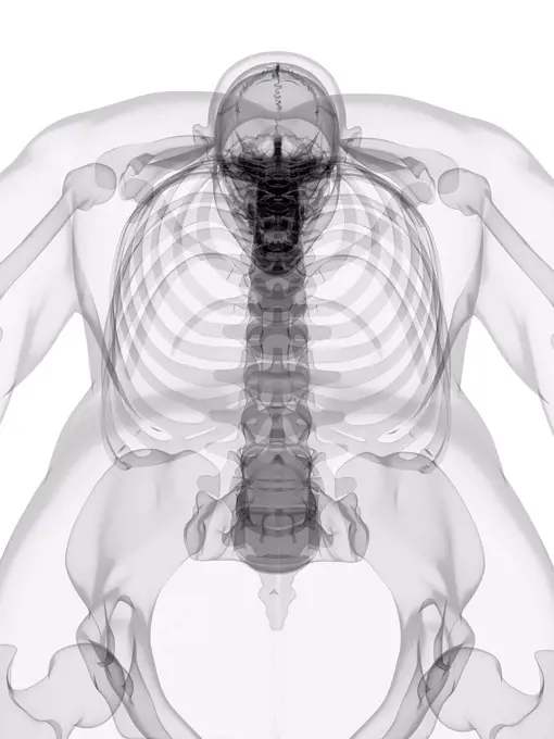
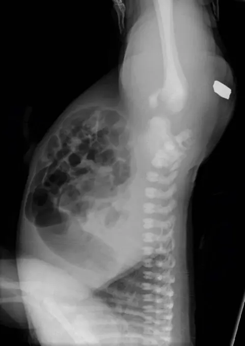
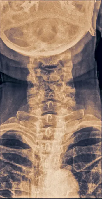
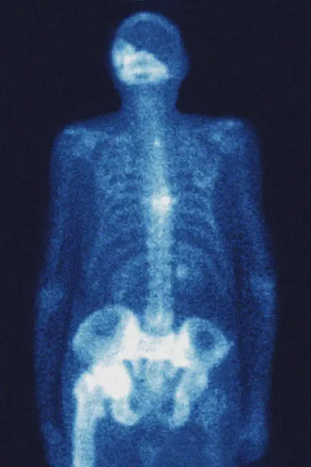
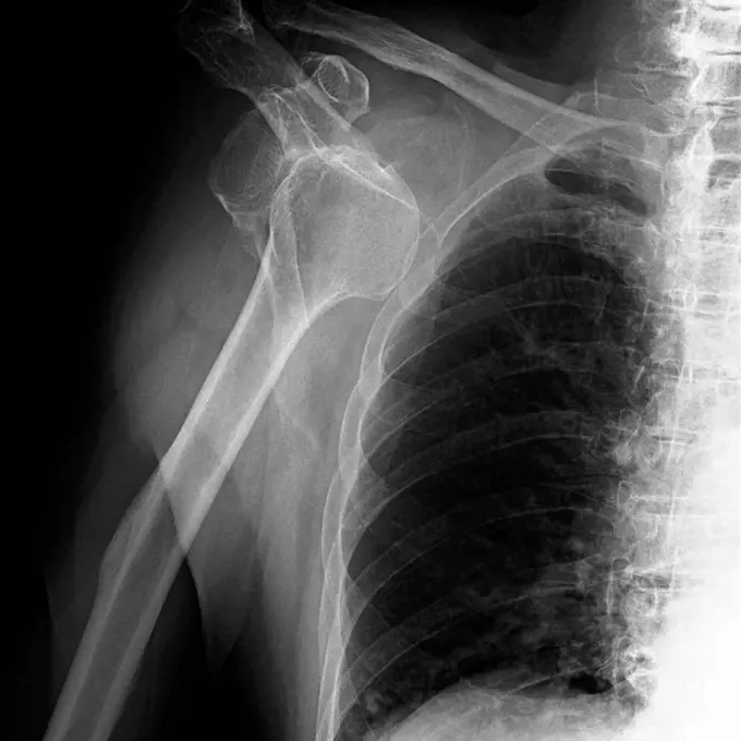
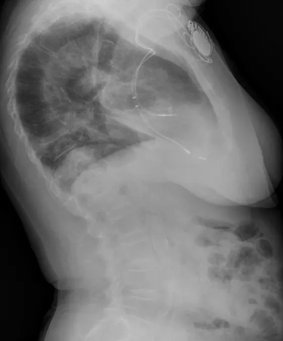
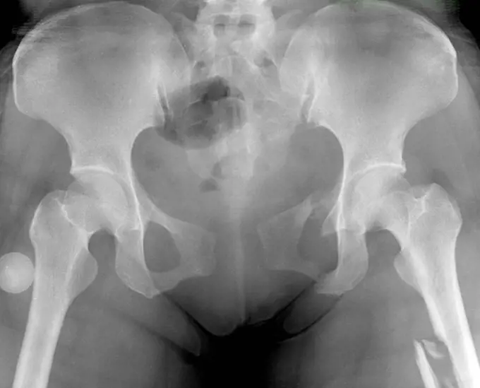
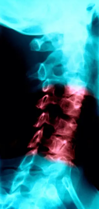
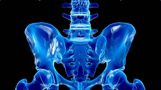
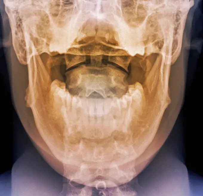
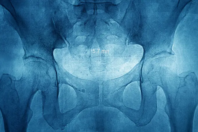
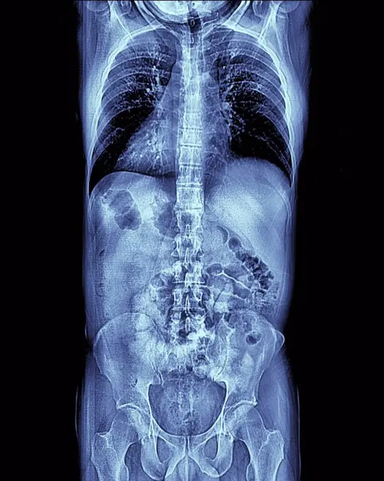
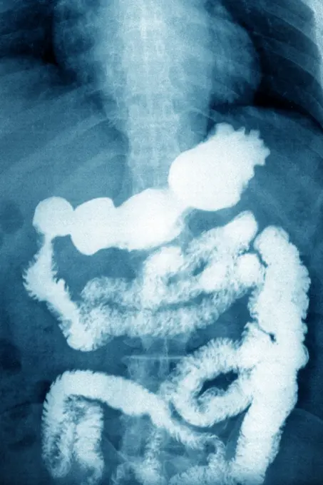
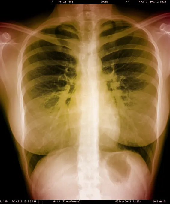
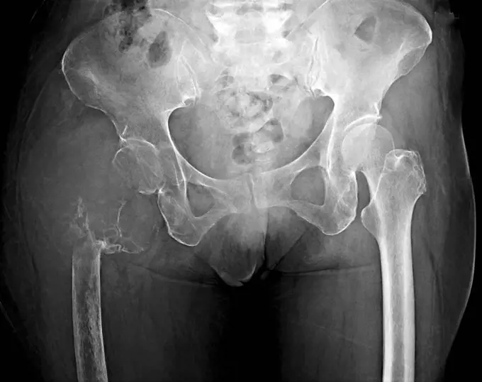
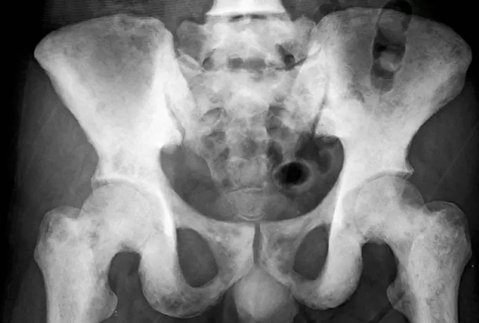
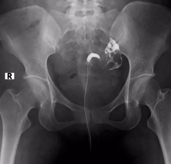
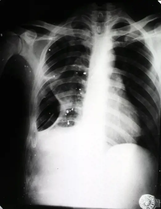
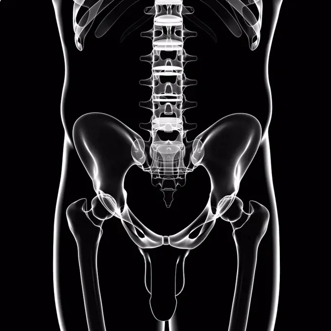
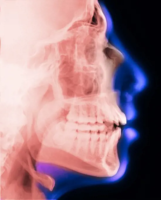
My Boards
View Pictures
  • My Boards

Spinal and Pelvic X-rays

X-ray images focusing on the human spine and pelvis, illustrating various medical conditions and anatomy in a clinical context.

X-ray styled view of the bones of the trunk.
235 assets in this story
4378-1941
X-Ray image showing the rib cage and pelvis.
4378-296
X-ray styled view of the bones of the trunk.
4128R-15536931
Illustration of the lumbar spine
5507-40438480
X-ray of the pelvis and spinal column
1815R-156186
Radiograph of lumbar spine
1899-54027389
X-ray of urinary schistosomiasis. Frontal pelvic x-ray. (Photo by: BSIP/UIG via Getty Images)
1815R-156185
Radiograph of lumbar spine
1899-53511888
Rotational dislocation at the junction of the thoracic and lumbar vertebrae, visualized by an AP chest radiograph. (Photo by: CAVALLINI JAMES/BSIP/UIG)
824-63226732
Rotational dislocation at the junction of the thoracic and lumbar vertebrae, visualized by an AP chest radiograph.
824-63128418
See image n0756005 on the site.
4128R-12660
Normal neck. Coloured X_ray of the cervical spine of a 27 year old.
824-63214248
X-ray of urinary schistosomiasis. Frontal abdominal-pelvic x-ray.
824-63175256
X-ray of the skull showing sinonasal polyposis.
1899-53511987
Chronic colitis (inflammatory bowel disease), revealed by a frontal abdominal x-ray.
6117-28548950
X-Ray Image Of Human Healthy Chest
824-63193990
A healthy lung x-ray on an 18-year old patient.
824-63204759
Frontal x-ray of a 60-year old woman's trunk.
824-63193987
A healthy lung x-ray on an 18-year old patient.
824-63193985
A healthy lung x-ray on an 18-year old patient.
4128R-5152
Normal lumbar spine, X_ray
824-63225320
Dislocation between the first and second cervical vertebrae, visualized by a sagittal x-ray of the cervical spine.
1439-57940808
Xray image revealing the human spine and pelvic bones.
1841R-111778
4128R-12677
Normal abdomen. X_ray of the abdomen of a 20 year old female.
4128R-1982
Normal human torso, monochrome profile magnetic resonance imaging MRI scan. The front of the body is at left. The spinal bones vertebrae are seen runn...
4128R-26344
Male skeleton, computer artwork.
824-63188625
An osteolytic-shaped malignant bone tumour in a lumbar vertebra, seen on a frontal x-ray.
4128-28970823
Invertogram X-ray
1439-57940360
Radiant xray image revealing the intricate bones of the human spine and pelvis.
1899-54027390
X-ray of urinary schistosomiasis. Frontal pelvic x-ray. (Photo by: BSIP/UIG via Getty Images)
1899-53511635
Paget's disease (bone involvement), visualized by scintigraphy.
824-63226763
Paget's disease (bone deformity of a thoracic vertebra), revealed by a sagittal radiograph of the thorax.
4128R-26251
Sacrum, computer artwork.
4128R-15536886
Illustration of the shoulder bones
6188-55478595
film with X-ray image of human female pelvis film with X-ray image of human female pelvis isolated on white background Copyright: xZoonar.com/ValeryxVoennyyx 13808323
1899-85843
Thorax X Ray
4269-27164
Thoracic outlet syndrome. Color enhanced 3D computed tomography (CT) scan showing a thoracic outlet syndrome (TOS).
1439-57940805
Xray image revealing the intricate structures within a human head.
824-63175258
X-ray of the skull showing sinonasal polyposis.
824-57655650
X-ray of spine affected by scoliosis, frontal view.
824-63214245
X-ray of urinary schistosomiasis. Frontal pelvic x-ray.
1525-26336671
X ray showing scoliosis of the lumbar spine. Scoliosis is an abnormal lateral curvature of the spine. Zoom in.
4128-30420004
Head and neck bones, X-ray
4128R-12774
Normal spine. Coloured X_ray of the spine of a 24 year old man.
1899-53511892
Dislocation of the cervical vertebrae, with spinal trauma, between the sixth and seventh cervical vertebrae, visualized by sagittal section radiography. (Photo by: CAVALLINI JAMES/BSIP/UIG)
4128R-36671
Male pelvic bones, computer artwork.
824-63184394
Sagittal plane scan of the right foot.
4128R-12678
Normal abdomen. Coloured X_ray of the abdomen of a 20 year old female.
824-63178577
X-ray of the pelvis in a 63-year-old patient suffering from pain in the joint of the left leg represented in red. No visible anomaly.
4269-25722
Hip osteoarthritis. X-ray showing arthritis of the hip. At lower left and right are the thigh bones., above is the pelvis. Arthritis is a joint disease that can cause cartilage destruction (seen as a reduction in the joint space, pink), bone erosions, and tendon inflammation.
1899-53511624
Dislocation between the first and second cervical vertebrae, visualized by a sagittal x-ray of the cervical spine.
4128R-2732
Normal torso, MRI scan
4128-19357524
X-ray of the pelvis of a patient who was in a traffic accident. The X-ray shows diastasis (separation) of symphysis pubis called symphysiolysis and a ...
4128-19358083
X-ray showing a dislocated shoulder, where the head of the humerus is detached from the shoulder joint.
1525-28007762
medical science image of human skeleton bones. Transparent Human Body with Visible Bones
1525-28468563
X ray film for human skull
4378-3702
Rear view of the anatomy of the back with transparent skin revealing the internal anatomy. Note the spinal anatomy is highlighted.
4378-3631
Front view of the torso anatomy with heart highlighted.
6145-29257524
Schistosomiasis: digestive system lesions
824-63194008
A healthy lung x-ray on an 18-year old patient.
824-63178593
X-ray of the pelvis and lower back of a 25 years old woman. Light discopathy on L4-L5 and L5-S1
1525-22610086
Neck and nape x-ray scan of a 42 year old adult male. Cervical vertebrae human anatomy.
4128-30421464
Posterior skeletal anatomy of the hip, illustration
4129-125
chicken pelvis x-ray
1574R-018937
X-ray of hips
4128R-12771
Normal shoulder. Coloured X_ray of the shoulder of a 19 year old man.
1899-66335
X-ray image of female chest, sternam, rib cage and spine
4128R-30959
Scoliosis of the spine. Coloured X-ray of the chest of a 31 year old patient with scoliosis (sideways curvature) of the spine.
4128-19358123
X-ray showing a dislocated shoulder, where the head of the humerus is detached from the shoulder joint.
1439-57940618
Medical chest radiograph Xray showing human thorax
255-28641405
Vintage Photograph. X Ray image of Cervical spine Lateral View
4128-19357533
X-ray of the pelvis of a patient who was in a traffic accident. The X-ray shows diastasis (separation) of the symphysis pubis called symphysiolysis an...
4128R-26338
Male skeleton, computer artwork.
1899-85878
Cervical Vertebra X Ray
4128-19056399
Sacrum, illustration
4128R-12668
Normal neck. Coloured frontal X_ray of the joint between the skull and the neck.
1525-24031592
3d illustration - x-ray panels on black background
824-63224749
Hip asymmetry, visualized by frontal pelvic x-ray.
824-63198847
Degenerative disk disease. Vertebral osteoarthritis seen on an side x-ray of the thorax.
4128R-2925
Normal human torso, monochrome frontal magnetic resonance imaging MRI scan. The spinal bones vertebrae are seen running from the top of the chest to t...
4128-30415769
Atlas bone, illustration
1899-66326
X-ray image of female lumbar vertibrae
824-63204760
Frontal x-ray of a 60-year old woman's trunk.
1899-54026884
Right and left ureter, seen on two frontal abdominal x-rays.
4128-30415797
Skeletal back, illustration
824-63123151
Normal.
824-63193991
A healthy lung x-ray on an 18-year old patient.
4269-25313
Prosthetic hip. X-ray of prosthetic hip showing the shaft of steel inserted into the femur (pink), with the artificial ball articulating with the bone socket of the pelvis.
824-63204708
Frontal x-ray exmaining gastrointentinal transit.
824-63194002
A healthy lung x-ray on an 18-year old patient.
4128-19357543
X-ray of the pelvis of a patient with aggressive primary bone tumor showing an ill-divined lytic lesion with wide zone transition, destruction of the ...
824-57655646
Profile x-ray of lungs.
4128-19357540
X-ray of the pelvis of a patient with prostate carcinoma. The X-ray shows an osteoblastic lesion on the pelvis bone and vertebra lumbalis indicating b...
4128-18929617
Malformed uterus, X-ray
1899-18977930
1944 - X-ray showing chest wound caused by a Japanese 50mm mortar shell.
824-63165990
Cervical vertebrae, 3D CT scan.
4128R-34300
Male pelvic bones, computer artwork.
4297-1378
Lateral skull x-ray of a 18 year old man
1525-25424267
X-ray of a human male chest
824-63226737
Dislocation of the cervical vertebrae, with spinal trauma, between the sixth and seventh cervical vertebrae, visualized by sagittal section radiography.
PREVIOUS
of 3
NEXT
2401 S. Ervay, Suite 206
Dallas, Texas 75215
United States
Get Started
Free ResearchMy BoardsMy Cart
For Creators
How To License Your ContentContributor PortalFrame of Mind
Resources
API accessPricing
Contact
+1 866 236 0087help@viewpictures.co.uk Contact form
©2026 View Pictures. All Rights Reserved. -A
This site is protected by reCAPTCHA and Google Privacy Policy and Terms of Service apply.