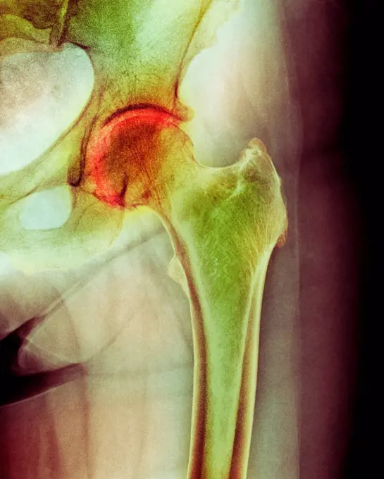
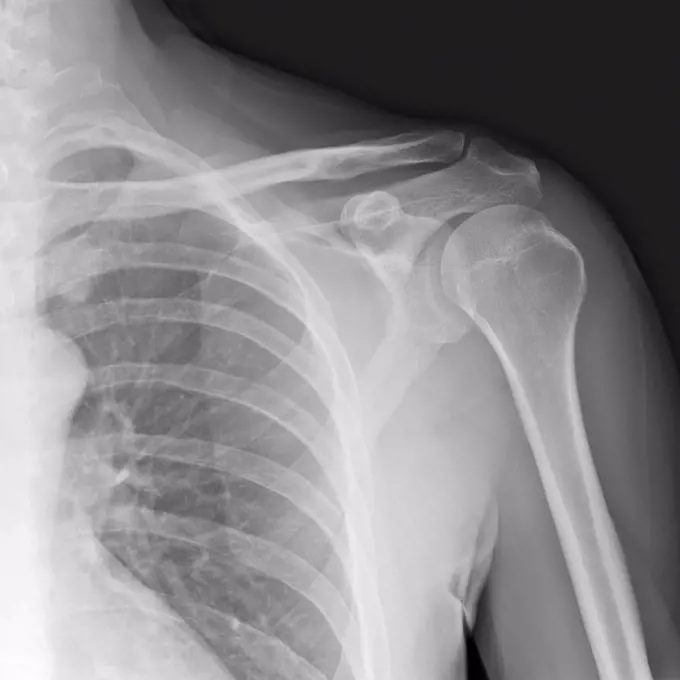
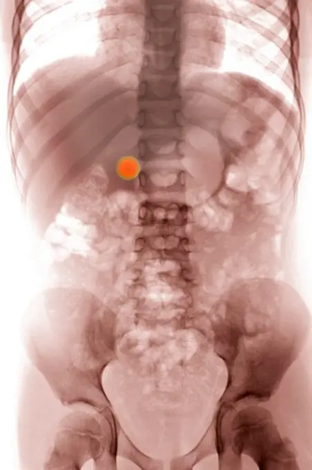
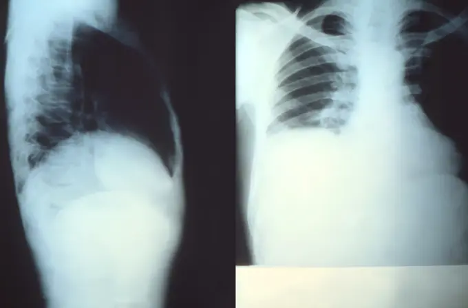
My Boards
View Pictures
  • My Boards

Spinal and Pelvic X-rays

X-ray images focusing on the human spine and pelvis, illustrating various medical conditions and anatomy in a clinical context.

X-ray styled view of the bones of the trunk.
235 assets in this story
4297-1076
Lumbar spine x-ray of a 41 year old man showing minor degenerative changes
1525-28468521
X ray film for human skull
4378-265
Close-up of stylized bones of the left hip within the skin.
824-63178554
X-ray of a child's sinuses. Normal
4269-24809
Disc prolapse. Colored X-ray of the lumbar rachis showing a narrowing of the intervertebral space in L5-S1 (in red), with an posterior osteophyte (abnormal bone production) extending on the lumber vertebral canal.
4269-25454
Hysterography. X-ray of female reproductive organs. A contrast medium was introduced into the uterus (center) and its Fallopian tubes (oviducts, thin, extending from uterus) through the uterine neck.
4269-27187
Human body. 3D Computed Tomography (CT) reconstruction scan of a human body revealing the skeleton, the heart, the kidneys (red), and the arterious system (yellow).
4128-19358120
X-ray of a 1 day old baby showing a normal chest, normal abdomen and bone structure.
4128-16375523
Oil drum skeleton, X-ray
4128-19358201
X-ray of a 1 day old baby showing a normal chest, normal abdomen and bone structure.
824-63188627
Bone metastases (ivory vertebrae) in a case of prostate cancer, seen on a saggital plane x-ray of the lumbar vertebrae.
1899-53508427
Aortic coarctation of newborn, before surgery, aortic coarctation is a narrowing of the aorta, often considerable and sometimes complete, most commonly just past the point where the aorta and the subclavian artery meet. Aortic coarctation causes low blood flow, often leading to arterial hypertension. Surgery is often advised to establish normal anatomy of the aortic intersection and gain normal blood flow. 3D CT scan.
824-63165968
Aortic coarctation of newborn, before surgery, aortic coarctation is a narrowing of the aorta, often considerable and sometimes complete, most commonly just past the point where the aorta and the subclavian artery meet. Aortic coarctation causes low blood flow, often leading to arterial hypertension. Surgery is often advised to establish normal anatomy of the aortic intersection and gain normal blood flow. 3D CT scan.
4128-24795785
Healthy stomach, barium X-ray
6188-55588298
Human teeth xray X ray of human mouth with teeth bones Copyright: xZoonar.com/ClaudioxDiviziax 8918967
1841R-111806
4128R-11323293
Human sacroiliac joint, computer artwork.
6188-63367372
X-Ray Image Of Human Chest - TB screening X-Ray Image Of Human Chest - TB screening. Copyright: xZoonar.com/GiovannixGagliardix 14522717
6145-52738563
X-ray image showing the knee joints of an Egyptian childs mummy from the Senckenberg Museum in Frankfurt, capturing bone structure and joint alignment, produced on bromide paper in 1896.
4128-19357514
X-ray of the lower (lumbarsacral) spine taken from a patient with ankylosis spondylitis and chronic lower back pain showing syndesmophytes.
6114-50976698
An x-ray of a fractured humerus.
1570R-132784
CAT Scan of a midsection of a person
4297-1141
CT scan images showing an abdominal aortic aneurysm which developed between the renal arteries and the common iliac arteries
4297-1037
Lateral view of the skull of a 43 year old woman without any teeth
1525-24739556
medical science image of human skeleton bones. Transparent Human Body with Visible Bones
4128R-15536938
Illustration of the sacrum
4128-28968805
Anatomy of the pelvis bones, illustration
4128R-12779
Normal hip. Coloured X_ray of the hip of a 90 year old man.
4128-28968808
Anatomy of the pelvis bones, illustration
6188-64760644
X-ray of a young three-year-old cat with bronchial asthma. Darkening of the bronchial type, interstitium without changes. A uniform decrease in the lumen of the trachea due to secondary collapse of the trachea. Excess amount of gas in the small intestine X-ray of a young three-year-old cat with bronchial asthma. Darkening of the bronchial type, interstitium without changes. A uniform decrease in the lumen of the trachea due to secondary collapse of the trachea. Excess amount of gas in the small intestine. Copyright: xZoonar.com/OlgaxOvchinnikovax 12143859
1525-26510343
Lungs ribs x-ray scan test result.
824-63197039
X-ray of the pelvis showing an intrauterine device (IUD).
6145-29296532
The bones of a shoulder joint and ribs, viewed through x-ray. Photoprint from radiograph after Sir Arthur Schuster, 1896.
824-63204764
X-ray of the whole spinal column, standing, from the front and side, with vertebrae angle measurements.
4128-30420595
Urinary system stent, X-ray
4297-1343
X-ray of the abdomen and pelvis of a man who came to the emergency room explaining that he had put some BBs into his penis before having had sexual intercourse with his female partner
1899-53508430
Large intercostal arteries, visible in a CT scan, developed to compensate pulmonary blood flow due to a congenital heart defect characterized by atresia of pulmonary arteries (small or absent pulmonary arteries). 3D CT scan.
824-63165971
Large intercostal arteries, visible in a CT scan, developed to compensate pulmonary blood flow due to a congenital heart defect characterized by atresia of pulmonary arteries (small or absent pulmonary arteries). 3D CT scan.
4128-18680909
Intussusception of the intestines, X-ray
1525-21265855
X-Ray pelvis shot isolated on white
4128-19358031
X-ray of a 1 day old baby showing a normal chest, normal abdomen and bone structure.
4269-27535
Pelvis fracture. 3D Computed Tomographic (CT) reconstruction scan of the pelvis showing a disjunction of the symphysis pubica (cartilage joining the two pubis). This trauma is frequently observed among victims of motorbike accidents.
4269-27534
Dislocation of the hip. 3D Computed Tomographic (CT) reconstruction scan of the pelvis showing a posterior hip luxation. The head of the femur (bone of the thigh) has totally slipped out on its insertion point  in the pelvis bone.
4378-183
Three-quarter stylized view of the bones of the left hip.
1815R-12499504
Pelvis x-ray
6188-55478592
film with X-ray image of human jaws isolated film with X-ray image of human jaws isolated on white background Copyright: xZoonar.com/ValeryxVoennyyx 13808297
1899-61460135
Spontaneous vertebral fracture due to oteoporosis (bone disease characterized by a decrease in bone mass, as well as bone deformation by compression and the risk of spontaneous fracture.). Lumbar vertebral x-ray
824-63225990
Lung cancer located in the upper right lobe. Frontal chest x-ray.
1899-66332
X-ray image of female wrist
4128-19489633
Colon diverticula, X-ray
4297-1342
normal shoulder x-ray of a 20 year old woman, showing the upper humerus and scapula
6114-50976625
An x-ray of a swallowed key in a childs stomach.
6145-52738440
The X-ray shows the right forearm of a female patient with a resection of one-third of the ulna due to caries from five years prior. Exposure time was 15 minutes. Printed on glossy bromide paper in a box format, 1896.
4128-19358080
X-ray showing a dislocated shoulder, where the head of the humerus is detached from the shoulder joint.
4128-30420028
Sinuses, X-ray
4220-21334635
Human Skeleton.     Date:
4269-5166
X-ray of a normal pelvis of a child.
824-33581
LUNG METASTASIS, X_RAY. Pulmonary metastases secondary cancer, carcinoma. Thoracic x_ray in front view.
4128R-12788
Arthritis of the hip. Coloured X_ray of an arthritic hip.
4356-319
An x-ray of a human shoulder from the front, showing the humerus (the upper arm bone), the shoulder blade or scapula, the collarbone or clavicle, and the rib cage.
1848-61113904
Magnetic resonance image of the lumbar spine
6145-46781585
Skeleton (front view).
1525-23488968
x ray picture of wild animal skeleton
5514-58777420
A collage of x-rays form a human skeleton.
4297-1196
Colorized x-ray of the abdomen of a 6 year old boy who swallowed a marble
824-57659367
This image depicts two thoracoabdominal x-rays, which reveal the presence of an enlarged liver, otherwise known as hepatomegaly. The x-ray on the left was taken from the right lateral perspective, and the x-ray at right was executed from the anteroposterior, or AP perspective. The enlargement of this patients liver was due to a parasitic infection known as amebiasis, caused by the protozoan organism, Entamoeba histolytica . In this case, youll note the dome of the enlarged liver, which is actually the diaphragm as it covers and curves over the superior surface of the liver, is at a higher level than it would normally be, encroaching upon the space within the thoracic cavity, which contains the heart and lungs. CDC/ Dr. Trenton K. Ruebush, III 1977
4269-27129
Kyphoplasty. Color enhanced sagittal computed tomography (CT) scan of the spine showing vertebral compaction due to osteoporosis after treatment by kyphoplasty.
4269-2536
X-ray showing an artificial implanted pancreas, a combination of a sensor that measures blood glucose level implanted in the superior vena cava (seen here), connected to an implanted insulin pump (not seen). Face view at left, and lateral view at right. The insuline is released automatically according the needs of the patient.
6145-29281958
Pelvis of E. Welch, showing disease of right hip and dislocaton of left hip. Radiograph, 1900/1904.
824-63178682
X-ray of the stomach of a young child. Transit without particular anomaly
6145-46781587
Skeleton (from behind).
4128-16375226
Pig skull, X-ray
824-63194028
X-ray of the collar bone and the left sternoclavicular joint. The sternoclavicular joint space seems a little pinched with a small subchondral osteosclerosis, degenerative in appearance. The exam shows a probable degenerative arthropathy of the sternoclavicular joint.
4128-19357553
X-ray of a pinned femur fracture with internal fixation plate and screw.
1899-66343
Dentistry X-ray image of female jaw and teeth
4128-19358082
X-ray showing a dislocated shoulder, where the head of the humerus is detached from the shoulder joint.
4128-19358081
X-ray showing a dislocated shoulder, where the head of the humerus is detached from the shoulder joint.
1848-55997721
X-ray of dog skull. Veterinary x-ray image
4128R-12785
Arthritis of the neck. Coloured X_ray of the arthritic cervical spine of a 70 year old man.
4297-1635
X-ray showing a spinal fusion of the lower lumbar vertebrae
824-63226759
Fixation by screws and plates on the sixth, seventh cervical vertebrae and the first thoracic vertebra, after vertebral dislocation, visualized by radiography in sagittal section.
1841R-111809
1841R-111802
6145-29288815
A skeleton of a new born child with deformed hips who died of asphyxia in birth. Collotype by Römmler & Jonas after a radiograph made for G. Leopold and Th. Leisewitz, 1908.
824-63188613
Osteoarthritis of the lumbar vertebrae seen on a side x-ray.
4128R-7297
Occluded intestine, X_ray
6145-29275356
A woman's pelvis after a succesful pubiotomy - a surgical incision of the symphysis pubis generally to widen the birth canal for vaginal delivery. Collotype by Römmler & Jonas after a radiograph made for G. Leopold and Th. Leisewitz, 1908.
824-57656904
Vertebral osteitis (inflammation of the bone tissue of a lumbar vertebra, caused by a bacterial infection). frontal x-ray of the lumbar vertebrae.
4128-30415795
Pelvis bone, illustration
1899-61459580
Röntgenaufnahme eines menschlichen Schädels, um 1915, Historisch, digital restaurierte Reproduktion von einer Vorlage aus dem 19. Jahrhundert.
5514-62181798
Microscopic view of a fetal chick skeleton.
4269-27144
Shoulder luxation. Colored X-ray showing a complete inferior dislocation of the shoulder. At left : normal position of the head of the humerus (upper arm bone in red) in the glenoid cavity (socket) of the scapula (shoulder blade). At right, the head of the humerus has slipped out backwards (inferior dislocation) of its socket in the shoulder blade.
1525-24739450
3d illustration of human digestive system parts and functions. human digestive system parts and functions
255-28641402
Vintage Photograph.  X-Ray image of an adult chest
824-738
Human Hip Showing Degenerative Joint Disease
1525-19826581
detail x-ray image scan of humans teeth
6145-45075038
The ridge in this image captured by NASA's 2001 Mars Odyssey spacecraft is the rim of a crater near the south polar cap.
824-642
X-ray image of a human foot
4128-16072984
Human hands on steering wheel, X-ray
1899-61460153
Vertebral osteitis (inflammation of the bone tissue of a lumbar vertebra, caused by a bacterial infection). frontal x-ray of the lumbar vertebrae
PREVIOUS
of 3
NEXT
2401 S. Ervay, Suite 206
Dallas, Texas 75215
United States
Get Started
Free ResearchMy BoardsMy Cart
For Creators
How To License Your ContentContributor PortalFrame of Mind
Resources
API accessPricing
Contact
+1 866 236 0087help@viewpictures.co.uk Contact form
©2026 View Pictures. All Rights Reserved. -B
This site is protected by reCAPTCHA and Google Privacy Policy and Terms of Service apply.