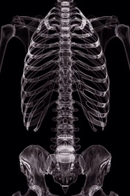
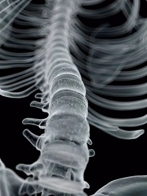
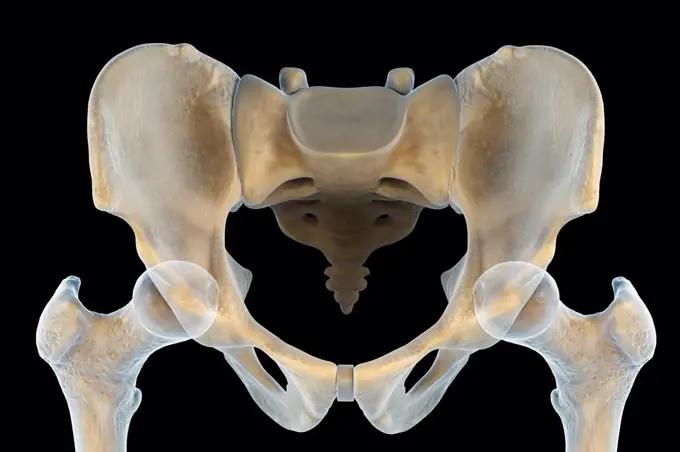
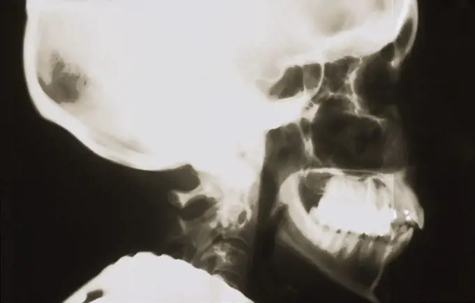
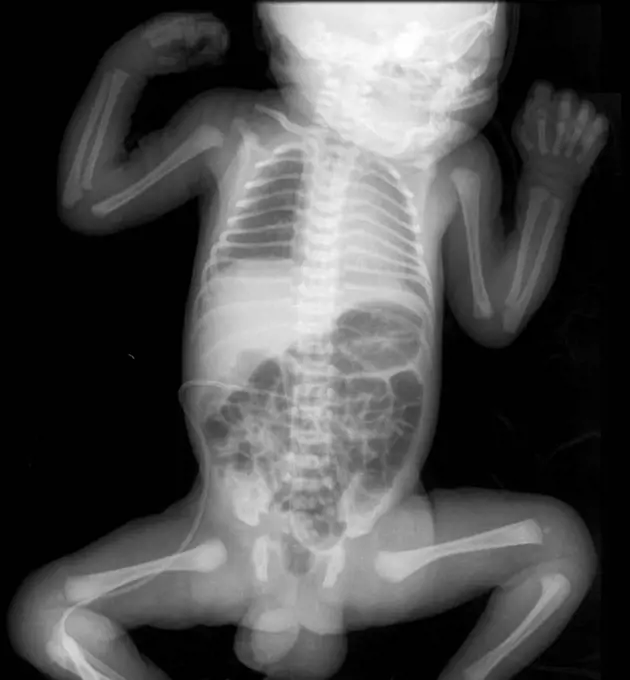
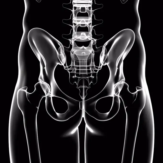
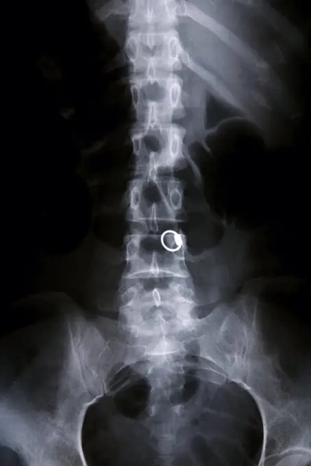
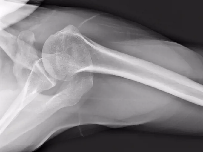
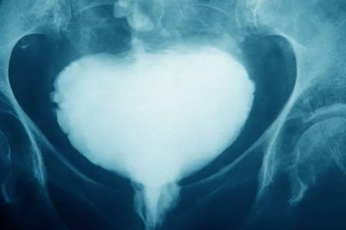
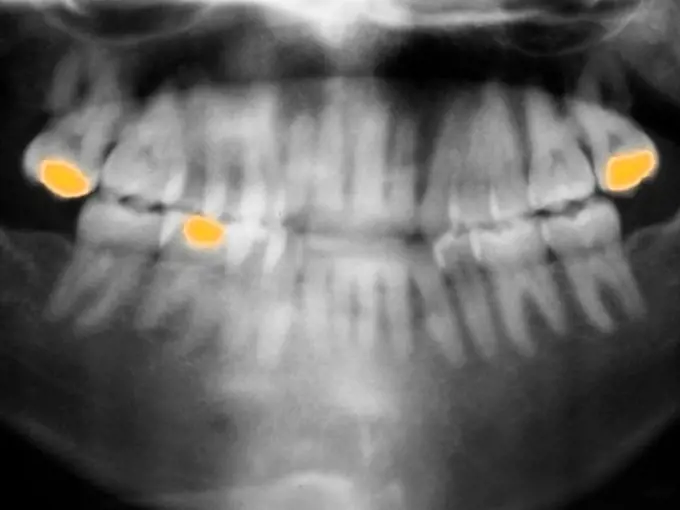
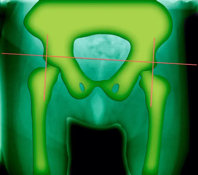
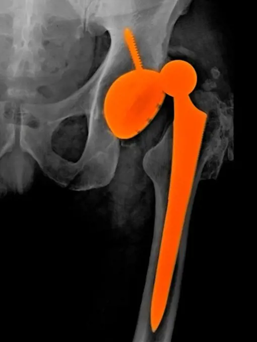
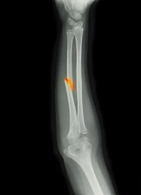
My Boards
View Pictures
  • My Boards

Spinal and Pelvic X-rays

X-ray images focusing on the human spine and pelvis, illustrating various medical conditions and anatomy in a clinical context.

X-ray styled view of the bones of the trunk.
235 assets in this story
824-63178715
Coloured x-ray of a ten years old boy dentition.
824-63208114
Osteoporosis with compression of the dorsal and lumbar vertebrae, seen on a sagittal plane MRI scan.
6145-52738552
The X-ray shows the supreme view of an Egyptian cat mummy from the Urban Historical Museum in Frankfurt am Main. The image is printed on glossy bromide paper in a box format from 1896.
824-63128419
The gallbladder is invaded by a cluster of small stones. The gallbladder is an oblong sac containing bile. It measures approximately 10 cm. It is located under the liver and therefore serves as a reservoir for the bile secreted by the latter. During the passage of the food bolus, it releases and evacuates the bile in the intestine, which allows the absorption of food and in particular fats. The gallbladder that we see here is the seat of a lithiasis (a lithiasis being a formation of stones). See image n0656805 on the site.
4128R-11324391
Human spine, computer artwork.
4378-3461
Front superior view of the male pelvis, sacrum and hip joints.
1525-22470463
an x-ray image the heart and lungs of a person shows.
1525-22143056
Xray of Skull
824-63224750
Hip asymmetry, visualized by frontal pelvic x-ray.
4128-19358215
X-ray of the chest showing righ pneumothorax (clear space in the right hemithorax).
4128-19358034
X-ray of a four day old baby showing a normal chest, normal abdomen and bone structure.
4128R-36537
Human pelvic bones, computer artwork.
1815R-6290
Radiograph of Cervical Spine
824-63194033
X-ray of the collar bone and the left sternoclavicular joint. The sternoclavicular joint space seems a little pinched with a small subchondral osteosclerosis, degenerative in appearance. The exam shows a probable degenerative arthropathy of the sternoclavicular joint.
4297-1204
Head x-ray of a 53 year old man with gunshot wounds
824-63194032
X-ray of the collar bone and the left sternoclavicular joint. The sternoclavicular joint space seems a little pinched with a small subchondral osteosclerosis, degenerative in appearance. The exam shows a probable degenerative arthropathy of the sternoclavicular joint.
6188-64760645
X-ray of a young three-year-old cat with bronchial asthma. Darkening of the bronchial type, interstitium without changes. A uniform decrease in the lumen of the trachea due to secondary collapse of the trachea. Excess amount of gas in the small intestine X-ray of a young three-year-old cat with bronchial asthma. Darkening of the bronchial type, interstitium without changes. A uniform decrease in the lumen of the trachea due to secondary collapse of the trachea. Excess amount of gas in the small intestine. Copyright: xZoonar.com/OlgaxOvchinnikovax 12143829
1841R-111818
4269-27145
Shoulder luxation. Colored X-ray showing a complete inferior dislocation of the shoulder. At left : normal position of the head of the humerus (upper arm bone in red) in the glenoid cavity (socket) of the scapula (shoulder blade). At right, the head of the humerus has slipped out backwards (inferior dislocation) of its socket in the shoulder blade.
824-63126492
3D image.
4356-322
An x-ray of a human shoulder from above, showing the humerus (the upper arm bone), and the shoulder blade or scapula.
1773R-87981
Model of human skull in lab
1899-54027408
Vesico-sigmoid fistulae. Frontal pelvic x-ray. (Photo by: BSIP/UIG via Getty Images)
824-63224672
Significant degenerative lumbar disc disease in an overweight 75-year-old lady. Lumbar CT scan in sagittal section.
6145-29260799
Anatomy: dissection of the thorax: the back. Photograph, ca. 1900.
4297-1085
Panorex x-ray of the jaw showing several fillings
1899-85984
Pelvis X Ray
4297-1059
X-ray of a 74 year old man with a dislocation of his hip replacement
1525-23488966
x ray picture of wild animal skeleton
6145-52282304
Aerial view of a part of the area affected during the flood disaster of 1953. Note: The description of this photo is not yet complete and will be supplemented on the basis of the data available at a later stage. However, of many photos of the flood disaster 1953, the location is unknown.
4269-38496
Colored X-ray showing the head of a man using a smartphone. The use of mobile phone generates microwaves that penetrate in the brain.
4128R-11287930
Dual chamber pacemaker. Coloured X-ray of the chest of a patient with a dual chamber pacemaker (top right). Wires lead from the device to both the rig...
4297-1600
Forearm x-ray of an 8 year old boy showing a fracture of the mid-shaft of the ulna
6145-46739447
Large copper mountain chronology. From Carl Sahlin's mountain historical collection.
6145-44735076
P29503 Moon (closeup) Voyager 2 flyby of Uranus
PREVIOUS
of 3
NEXT
2401 S. Ervay, Suite 206
Dallas, Texas 75215
United States
Get Started
Free ResearchMy BoardsMy Cart
For Creators
How To License Your ContentContributor PortalFrame of Mind
Resources
API accessPricing
Contact
+1 866 236 0087help@viewpictures.co.uk Contact form
©2026 View Pictures. All Rights Reserved. -A
This site is protected by reCAPTCHA and Google Privacy Policy and Terms of Service apply.