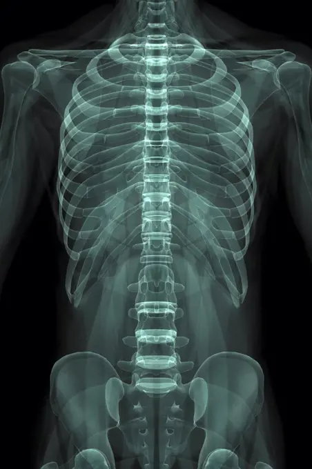
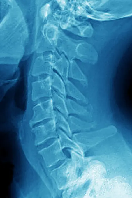
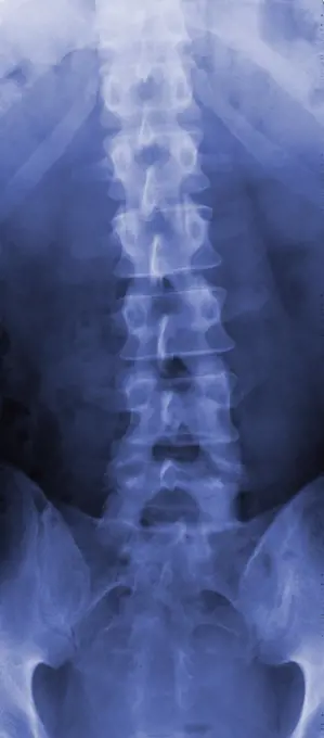
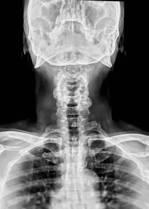
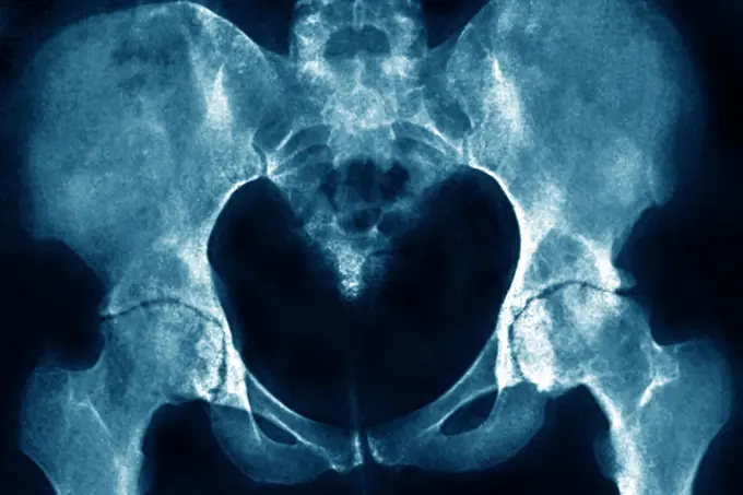
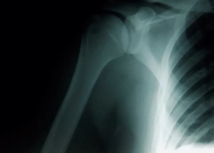
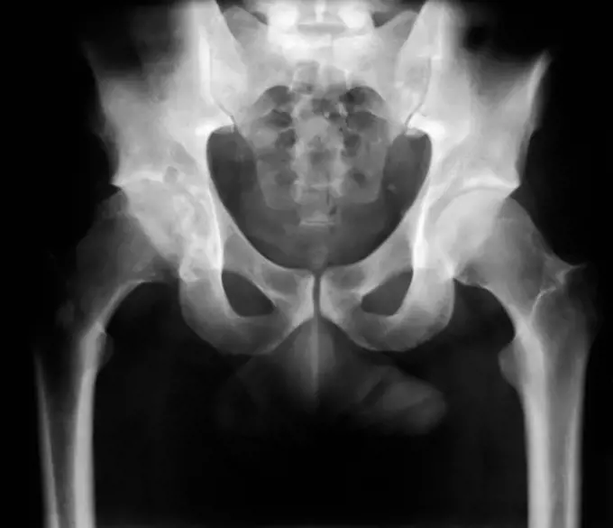
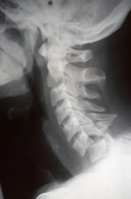
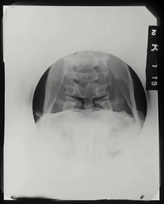
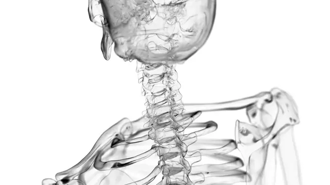
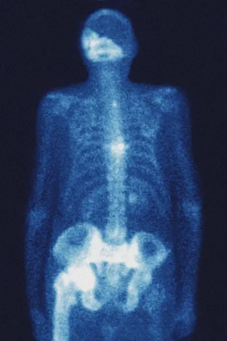
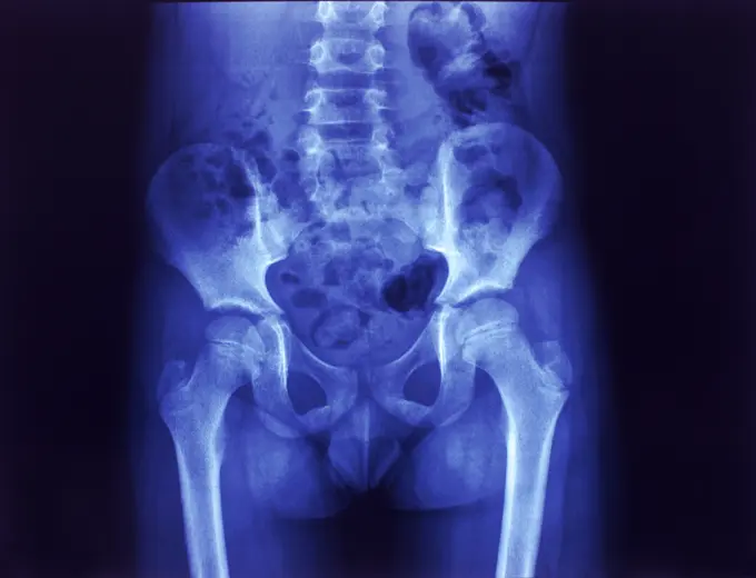
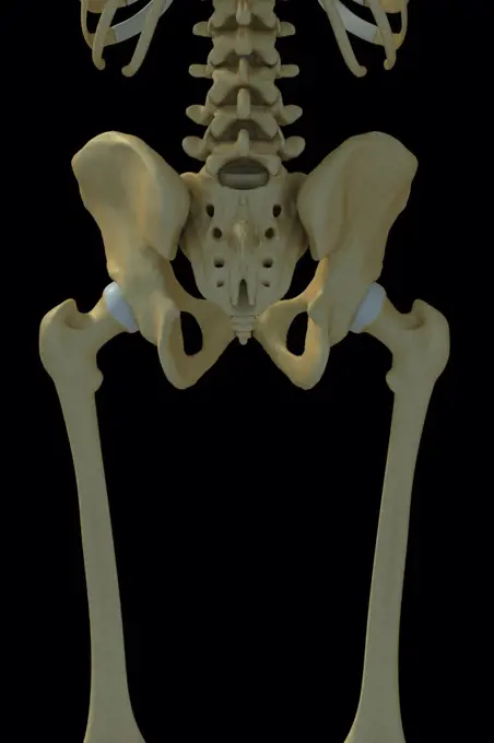
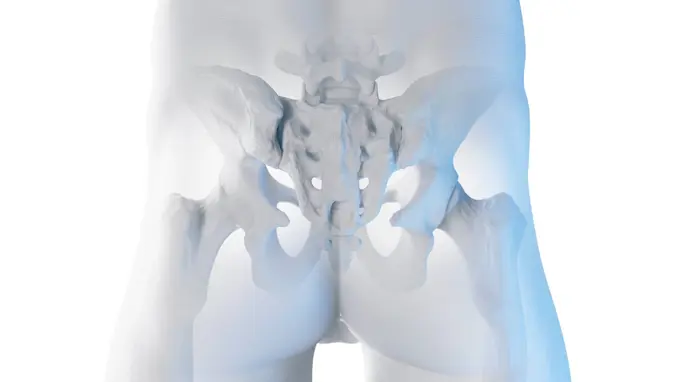
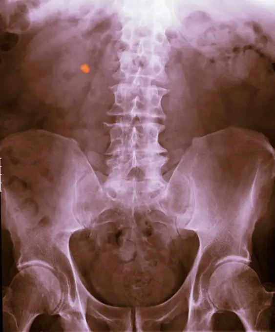
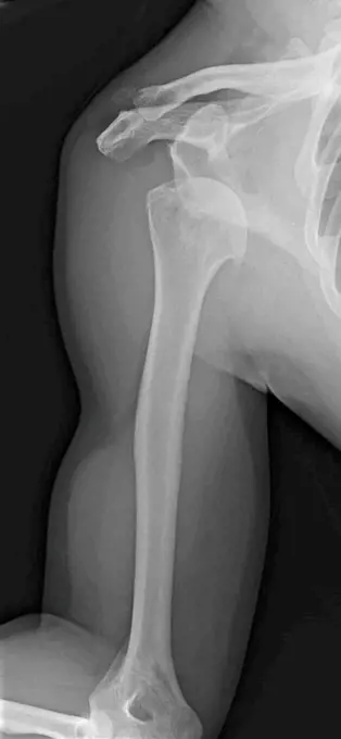
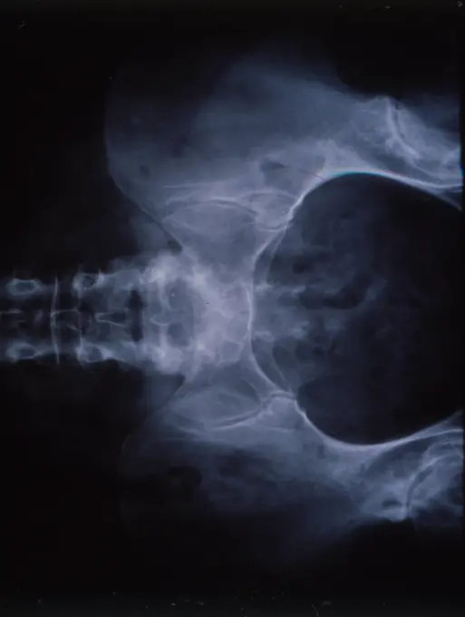
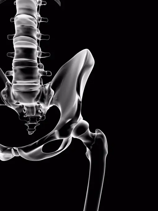
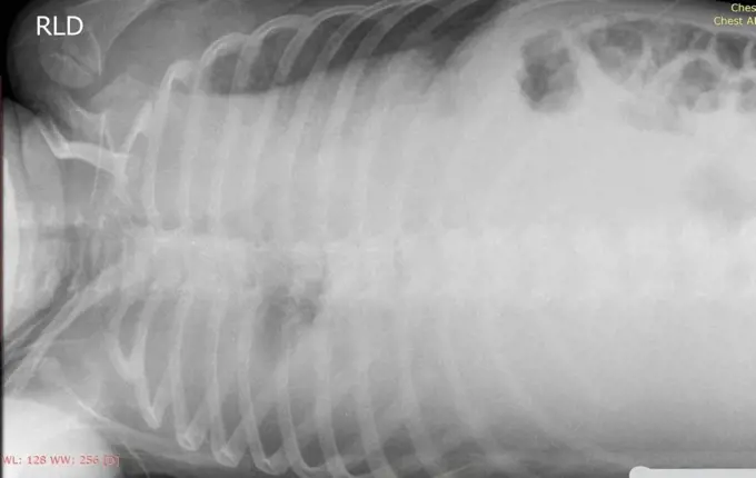
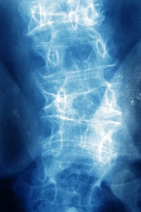
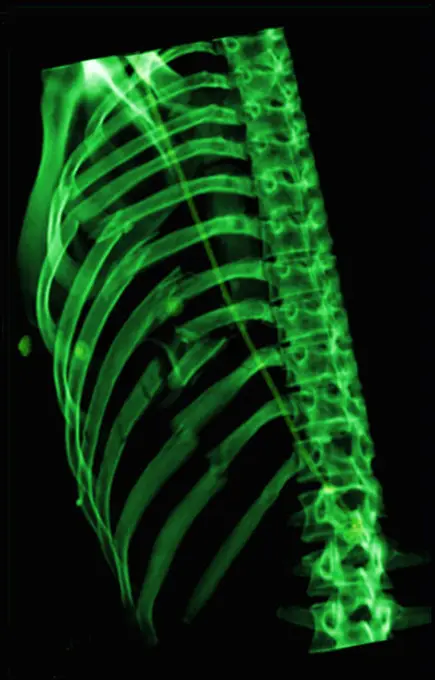
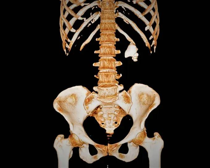
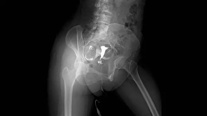
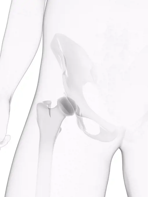
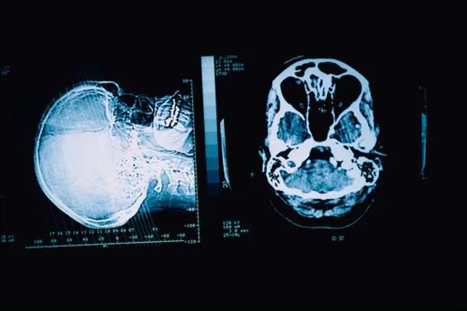
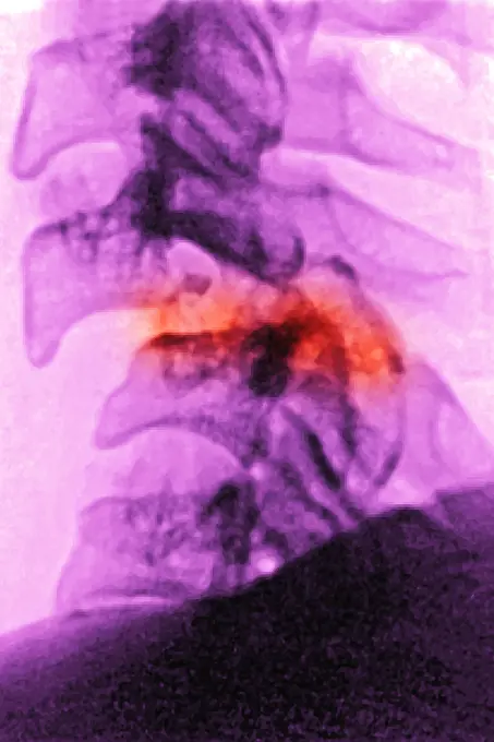
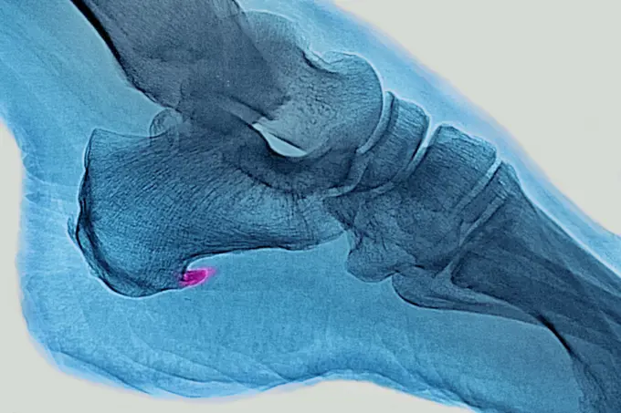
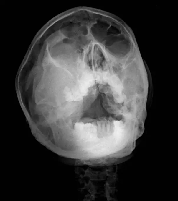
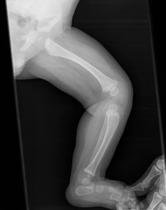
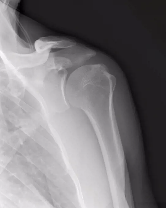
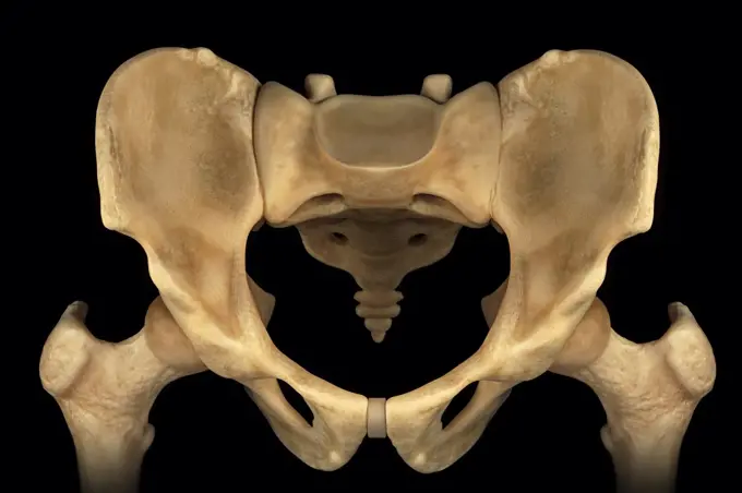
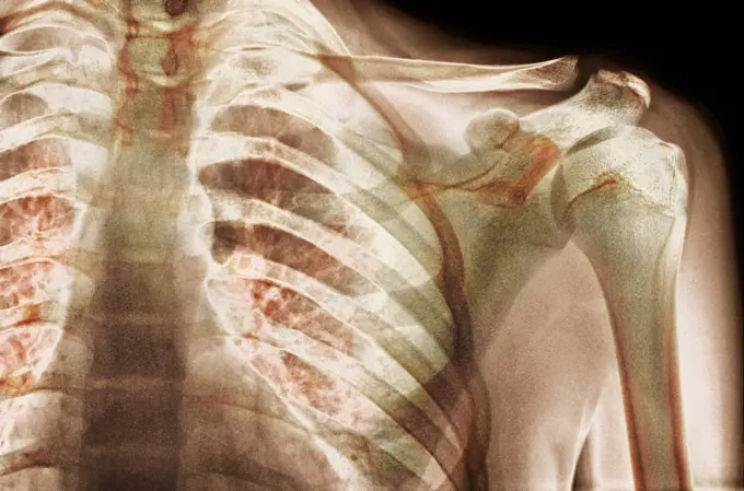
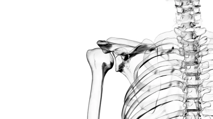
My Boards
View Pictures
  • My Boards
← Science

X-Ray Images of the Spine and Torso

A collection of x-ray images illustrating the spinal column, rib cage, and pelvic area. Highlights conditions such as osteoarthritis with a clinical focus.

X-Ray image showing the rib cage and pelvis.
150 assets in this story
1990-12808
xray of torso
824-63225966
Cervical osteoarthritis visualized by a sagittal x-ray of the neck.
4378-1945
X-Ray image showing the rib cage and pelvis.
1841R-111400
radiograph of a spinal column
4128R-12683
Normal extended neck. Coloured X_ray of the extended cervical spine of a 20 year old female.
4128-30420005
Head and neck bones, X-ray
4128R-12669
Normal neck. X_ray of the cervical spine of a 24 year old.
1899-53511741
Osteoarthritis of the hips (coxarthrosis) visualized by a pelvic x-ray of the front. (Photo by: CAVALLINI JAMES/BSIP/UIG)
4128R-12679
Normal abdomen. Coloured X_ray of the abdomen of a 20 year old female.
4128R-15536946
Illustration of the sacrum
1848-49284873
X ray vertebral column
1525-22924372
3D magnetic resonance image scan
1525-21748212
detail x-ray image scan of humans teeth
6188-55478590
human jaws on X-ray image front view of human jaws on X-ray image Copyright: xZoonar.com/ValeryxVoennyyx 13808244
255-28641412
Vintage Photograph. X Ray image of Cervical Spine, Lateral View
1525-19848582
detail x-ray image scan of humans teeth
4128R-15536816
Illustration of the cervical spine
6145-29287106
The bones of a knee joint of a boy, viewed through x-ray. Photoprint from radiograph after Sir Arthur Schuster, 1896.
824-63178586
X-ray of the sinuses of a 35-year-old woman. Hypertrophy of the right and left middle and inferior turbinates
1899-66327
X-ray image of female clavical, scapula and upper humerous
4362-749
X-Ray of Human Male Pelvis
1899-53511508
Hip asymmetry, visualized by frontal pelvic x-ray.
1841R-111795
1525-22403807
Profile xray of a skull.
1525-19825169
Panoramic x-ray image scan of humans teeth
6114-50976642
An x-ray of a dislocated cervical spine.
4378-3714
The bones of the male pelvis viewed from the front.
4378-2988
Wire frame anatomical model showing the sternoclavicular articulation.
255-28641404
Vintage Photograph. X Ray image of the Anterior posterior view of first two vertebrae of Cervicle spine through open mouth
4128-19056347
Neck joint, illustration
824-63225333
Paget's disease (bone involvement), visualized by scintigraphy.
4378-3580
Rear view of the male pelvis, sacrum and hip joints.
1525R-194553
Profile xray of a skull.
4297-1106
Normal lateral skull x-ray of a 2 year old boy
4128-28769224
Sinus pain, conceptual X-ray
6188-58102441
Human Body with Visible Skeletal Bones science anatomy scan of human body with visible skeletal bones Copyright: xZoonar.com/StanislavxRishnyakx 12860675
6188-58104061
Transparent Human Body with Visible Bones medical science image of human skeleton bones Copyright: xZoonar.com/StanislavxRishnyakx 13861314
4128R-12964633
Human hip bone, Illustration
4269-5167
X-ray of a normal pelvis of a child.
1439-57940806
An Xray image of a human skull in monochrome.
1899-54027419
Vesicule calculus. Frontal pelvic x-ray. (Photo by: BSIP/UIG via Getty Images)
824-63178595
X-ray of the pelvis and lower back of a 25 years old woman. Light discopathy on L4-L5 and L5-S1
1899-66341
X- ray of lower back vertibrae
4128R-11324438
Human hip bone, computer artwork.
824-63195601
Abdominal x-ray showing an adenocarcinoma of the intestine ( neoplastic colic ferrule).
4128R-26343
Male skeleton, computer artwork.
4128R-12580464
Human spine, computer illustration.
4378-1950
X-ray image showing the shoulder joint.
4378-499
Rear view of the bones of the upper legs and pelvis.
4378-2077
X-ray showing the skeletal structure of the human pelvis.
4128-30421455
Posterior skeletal anatomy of the hip, illustration
4297-1064
Abdomen x-ray of a 73 year old man with a 1 cm stone in his right kidney
4128-19358217
X-ray showing a dislocated shoulder, where the head of the humerus is detached from the shoulder joint.
6188-55645577
X -ray of the pelvis and the spine X -ray image of the pelvis and the spine
824-63225957
Right coronary artery visualized by frontal thoracic angiography.
4128R-11310860
Human pelvis, computer artwork.
4128-20043642
Pleural effusion, X-ray
824-65830292
Lumbar osteoporosis (disease causing compression and fractures of the lumbar vertebrae). X-ray of the lumbar vertebrae seen from the front.
4269-27512
Costal fracture. 3D Computed Tomography (CT) reconstruction scan of the thoracic cage showing multiple fractured ribs.
4128-30420026
Human skull, X-ray
1558-14163610
X-ray picture of a human bite,
4128-48285509
Kidney stone, 3D CT scan
1525-22310734
Close-up of a dental X-Ray
6177-V53850465
4128R-30117
Male pelvis bones, computer artwork.
4128-20041867
Fallopian tube of a patient with infertility, X-ray
824-57656877
Spontaneous vertebral fracture due to oteoporosis (bone disease characterized by a decrease in bone mass, as well as bone deformation by compression and the risk of spontaneous fracture.). Lumbar vertebral x-ray.
824-63208110
Osteoporosis with compression of L1-L2 vertebrae, seen on a sagittal plane MRI scan.
6188-55645093
Abdomen man, man abdomen man, man, ct recording copyright: xzoonar.com/dr.xnorbertxlangeex 13790167
4128R-30118
Male pelvis bones, computer artwork.
1525-22787181
science anatomy scan of human body with visible skeletal bones. Human Body with Visible Skeletal Bones
1848-55997728
X-ray film of dog lateral view. Veterinary medicine, veterinary anatomy Concept
6096-19418399
"If you saw us" # 1. Report on the nights of young people in Bretagne. "Si l'on nous voyait" #1. Reportage sur les nuits des jeunes en Bretagne.
4286-39878
CT scans of a human skull and brain.
1899-53511893
Dislocation of the cervical vertebrae, with spinal trauma, between the sixth and seventh cervical vertebrae, visualized by sagittal section radiography. (Photo by: CAVALLINI JAMES/BSIP/UIG)
824-63212843
Enthesopathy: calcifications of the plantar fascia. Visualisation on saggital plane x-ray.
1848-62615589
X-Ray of a goshawk wing with a splint
6188-56014173
X-Ray of the pelvis and both hips of a little boy X-Ray of the pelvis and both hips of a little boy, a horizontal picture Copyright: xZoonar.com/OlgaxPopovax 3619728
4128-24795479
Maxillary mass, X-ray
4128-19248773
Spine, illustration.
1439-57940797
Xray image showing a bent knee joint
824-63194031
X-ray of the collar bone and the left sternoclavicular joint. The sternoclavicular joint space seems a little pinched with a small subchondral osteosclerosis, degenerative in appearance. The exam shows a probable degenerative arthropathy of the sternoclavicular joint.
4128R-11478170
Human pelvis rendered in glass, computer artwork.
4356-320
An x-ray of a human shoulder from behind, showing the humerus (the upper arm bone), and the shoulder blade or scapula.
4128R-31661
Female skeleton, computer artwork.
1899-53508440
Pelvis and iliac arteries are visible, as well as a stent in the gluteal artery. 3D CT scan.
4269-25092
Fractured femur neck. Colored X-ray of the right hip of a patient, showing a fracture of the neck of the femur (yellow). The femoral head (round, center left) has separated from the femoral shaft (lower left) due to this fracture. Osteoporosis can cause this type of fracture.
4378-3958
Front superior view of the male pelvis, sacrum and hip joints.
6145-59512532
X -ray of the tbc -infected spine of Isabel Wachenheimer, dated 16 June 1948, Anonymous, 1948 photograph X -ray of the tbc -infected vertebra of Isabel Wachenheimer.  photographic support gelatin silver print
4128R-12772
Normal shoulder. Coloured X_ray of the shoulder of a 19 year old man.
4378-3596
The bones of the male pelvis viewed from a three-quarter perspective.
1899-53511511
Hip asymmetry, visualized by frontal pelvic x-ray.
6145-46697050
Coose search with X-rays.Radiografi of Lecturer Zallander's hand, taken in December 1896. Exposure time: 15 min with Crook's tube. Electricity source: Wimsherst's hand-driven flu machine.
4378-4073
Cervical Vertebrae
824-63178588
X-ray of the sinuses of a 35-year-old woman. Hypertrophy of the right and left middle and inferior turbinates
4269-27533
Dislocation of the hip. 3D Computed Tomographic (CT) reconstruction scan of the pelvis showing a posterior hip luxation. The head of the femur (bone of the thigh) has totally slipped out on its insertion point  in the pelvis bone.
4128R-12580708
Human nerves, computer illustration.
4128R-12964643
Human skeletal system, Illustration
4269-27532
Pelvis luxation. 3D Computed Tomographic (CT) reconstruction scan of the pelvis showing a luxation of the symphysis pubica (cartilage joining the two pubis) further to a compaction.
4128-19056439
Shoulder joint, illustration
PREVIOUS
of 2
NEXT
2401 S. Ervay, Suite 206
Dallas, Texas 75215
United States
Get Started
Free ResearchMy BoardsMy Cart
For Creators
How To License Your ContentContributor PortalFrame of Mind
Resources
API accessPricing
Contact
+1 866 236 0087help@viewpictures.co.uk Contact form
©2026 View Pictures. All Rights Reserved. -A
This site is protected by reCAPTCHA and Google Privacy Policy and Terms of Service apply.