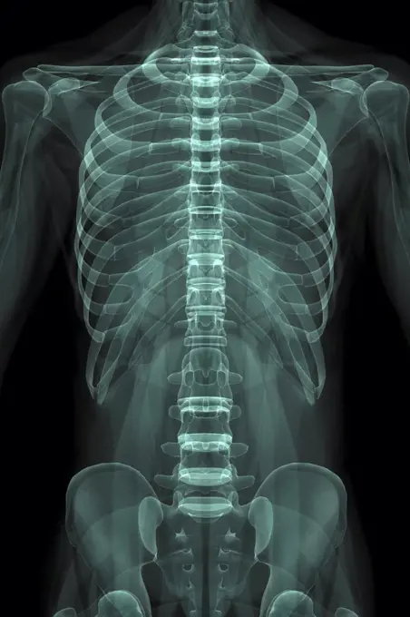
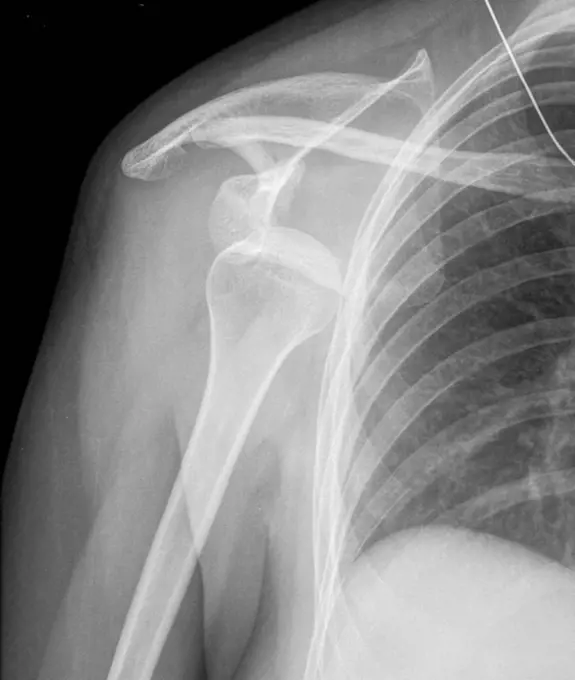
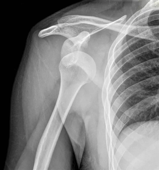
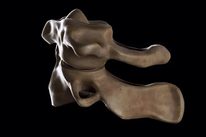
X-Ray Images of the Spine and Torso

A collection of x-ray images illustrating the spinal column, rib cage, and pelvic area. Highlights conditions such as osteoarthritis with a clinical focus.

X-Ray image showing the rib cage and pelvis.
Coloured x-ray of a ten years old boy dentition.Human teeth xrayRibs and vertebras. 3D computed tomographic (CT) scan reconstruction showing the middle part of the backbone and the lower ribs seen from behind.X-ray showing a dislocated shoulder, where the head of the humerus is detached from the shoulder joint.Anatomy of the coxal bones, illustrationRibs and vertebras. 3D computed tomographic (CT) scan reconstruction showing the middle part of the backbone and the lower ribs (side view).Kyphoplasty. Colored X-ray of the spine of a patient with vertebral compaction due to osteoporosis treated by kyphoplasty. The erosion of the bone structure of the vertebrae causes its deformation. Balloons are first inserted in the body of vertebrae and inflated in order to restaure its normal size. Surgical cement is then injected in the body (dark blue).X-ray showing a dislocated shoulder, where the head of the humerus is detached from the shoulder joint.3D CT scan of stenosed carotid artery, responsable for stroke.Vertebral osteoarthritis with osteophytes (bone spurs) seen on a saggital plane x-ray of the thoracic vertebrae.Osteosynthesis. 3D computed tomographic (CT) scan reconstruction (side view) of the lower backbone showing the sacrum and last lumbar vertebras (L4 and L5) maintained by pins further to a fracture.Human scapula and humerus, computer artwork.radiographic scan of homan skeletal bonesThe bones of the ribs and vertebral column, viewed through x-ray. Photoprint from radiograph after Sir Arthur Schuster, 1896.Osteoporosis of the lumbar vertebrae, seen on a side x-ray.X-ray photography taken by Oscar Karlowitch Larsson at Åminne, Mount or Sockholm.Atlas and Axis VertebraeX-ray of the collar bone and the left sternoclavicular joint. The sternoclavicular joint space seems a little pinched with a small subchondral osteosclerosis, degenerative in appearance. The exam shows a probable degenerative arthropathy of the sternoclavicular joint.Enlarged heart, X_rayIllustration of a man's larynxThree-dimensional computed tomographic reconstruction of pregnant woman pelvis showing the skeleton of a 9 months old fetus, pelvimetry allows to measure the diameter of a woman at the 9th month of pregnancy.Human femur head, computer artwork.Cervical spine, computer artwork.Edited illustrationAnatomy of the Spine on black background 3D illustration. Anatomy of the Spine on black backgroundX-ray showing a dislocated shoulder, where the head of the humerus is detached from the shoulder joint.X-ray of the stomach of a young child. Transit without particular anomalyHuman heel bones, IllustrationX-ray of the stomach of a young child. Transit without particular anomalyShoulder joint, MRISpine, illustration.Prostate cancer seen by frontal pelvic x-ray. (Photo by: CAVALLINI JAMES/BSIP/UIG)Halitherium Schinzi, vintage engraved illustration. From the Universe and Humanity, 1910.Chicken leg, X-rayKnee joint, illustrationSkeletal structure, IllustrationAnatomy of the Spine on black background 3D illustration. Anatomy of the Spine on black backgroundVertebral osteoarthritis with osteophytes (bone spurs) seen on a saggital plane x-ray of the thoracic vertebrae.Seattle, Washington:  1962. The University of Washingon crewman work out on Lake Washington in their shell.dental X-ray picture of human jaws isolated dental X-ray picture of human jaws isolated on white background ,property released Copyright: xZoonar.com/ValeryxVoennyyx 7103484 ,model released, Symbolfoto ,property releasedCAT scan of a skullHuman foot seen on a 3D CT scan.Cattle anatomy, illustration.Ram skull with horns, X-rayAnatomy: a dissection of the thorax: the thorax wall. Photograph, ca. 1900.Normal spine. Coloured X_ray of the spine of a 24 year old man.3D reconstruction from a CT image of a patient's jaw for personalized 3D printing.Colored X-ray showing the head of a man using a smartphone. The use of mobile phone generates microwaves that penetrate in the brain.3d illustration - x-ray panels on black backgroundRoentgenbild der Zaehne, Panoramaaufnahme der gesamten Zaehne im Ober- und Unterkiefer X-ray of the teeth, panoramic image of the entire teeth in the upper and lower jaw BLWS718051 *** X-ray image the Teeth, Panorama photo the entire Teeth at From and Lower jaw X Ray of The teeth, Panoramic Image of The Entire Teeth in The Upper and Lower JAW BLWS718051 Copyright: xblickwinkel/fotototox